 Movie
Movie Controller

[English] 日本語
 Yorodumi
- PDB-4lno: B. subtilis glutamine synthetase structures reveal large active s... -
+ Open data

- Basic information
| Entry | Database: PDB / ID: 4lno | ||||||
|---|---|---|---|---|---|---|---|
| Title | B. subtilis glutamine synthetase structures reveal large active site conformational changes and basis for isoenzyme specific regulation: form two of GS-1 | ||||||
 Components Components | Glutamine synthetase | ||||||
 Keywords Keywords | LIGASE / alpha/beta / tnra / glnr | ||||||
| Function / homology |  Function and homology information Function and homology informationcellular response to nitrogen levels / L-glutamine binding / nitrogen catabolite repression of transcription / glutamine synthetase / : / glutamine synthetase activity / glutamate binding / transcription regulator inhibitor activity / DNA-binding transcription factor binding / negative regulation of DNA-templated transcription ...cellular response to nitrogen levels / L-glutamine binding / nitrogen catabolite repression of transcription / glutamine synthetase / : / glutamine synthetase activity / glutamate binding / transcription regulator inhibitor activity / DNA-binding transcription factor binding / negative regulation of DNA-templated transcription / magnesium ion binding / ATP binding / cytoplasm Similarity search - Function | ||||||
| Biological species |  | ||||||
| Method |  X-RAY DIFFRACTION / SYNCHROTRON / MOLECULAR REPLACEMENT / Resolution: 2.9 Å X-RAY DIFFRACTION / SYNCHROTRON / MOLECULAR REPLACEMENT / Resolution: 2.9 Å | ||||||
 Authors Authors | Schumacher, M.A. / Chinnam, N. / Tonthat, N. / Fisher, S. / Wray, L. | ||||||
 Citation Citation | Journal: J.Biol.Chem. / Year: 2013 Title: Structures of the Bacillus subtilis Glutamine Synthetase Dodecamer Reveal Large Intersubunit Catalytic Conformational Changes Linked to a Unique Feedback Inhibition Mechanism. Authors: Murray, D.S. / Chinnam, N. / Tonthat, N.K. / Whitfill, T. / Wray, L.V. / Fisher, S.H. / Schumacher, M.A. | ||||||
| History |
|
- Structure visualization
| Structure viewer | Molecule: MolmilJmol/JSmol |
|---|
- Downloads & links
-Download
| PDBx/mmCIF format | 4lno.cif.gz | 534.6 KB | Display | PDBx/mmCIF format |
|---|---|---|---|---|
| PDB format | pdb4lno.ent.gz | 439.9 KB | Display | PDB format |
| PDBx/mmJSON format | 4lno.json.gz | Tree view | PDBx/mmJSON format | |
| Others |  Other downloads Other downloads |
-Validation report
| Arichive directory | https://data.pdbj.org/pub/pdb/validation_reports/ln/4lnoftp://data.pdbj.org/pub/pdb/validation_reports/ln/4lno | HTTPS FTP |
|---|
-Related structure data
| Related structure data | 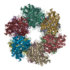 4lnfC 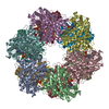 4lniC 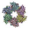 4lnkC 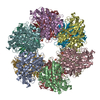 4lnnC 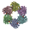 2bvcS C: citing same article ( S: Starting model for refinement |
|---|---|
| Similar structure data |


-Links
 PDBj
PDBj
- Assembly
| Deposited unit | 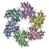
| ||||||||
|---|---|---|---|---|---|---|---|---|---|
| 1 | 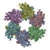
| ||||||||
| Unit cell |
|
-Components
| #1: Protein | Mass: 50206.906 Da / Num. of mol.: 6 Source method: isolated from a genetically manipulated source Source: (gene. exp.) #2: Chemical | ChemComp-MG /   Mass: 24.305 Da / Num. of mol.: 12 / Source method: obtained synthetically / Formula: Mg Mass: 24.305 Da / Num. of mol.: 12 / Source method: obtained synthetically / Formula: Mg#3: Chemical | ChemComp-GLN /   Type: L-peptide linking / Mass: 146.144 Da / Num. of mol.: 6 / Source method: obtained synthetically / Formula: C5H10N2O3 Type: L-peptide linking / Mass: 146.144 Da / Num. of mol.: 6 / Source method: obtained synthetically / Formula: C5H10N2O3#4: Water | ChemComp-HOH / |  Mass: 18.015 Da / Num. of mol.: 713 / Source method: isolated from a natural source / Formula: H2O Mass: 18.015 Da / Num. of mol.: 713 / Source method: isolated from a natural source / Formula: H2O |
|---|
-Experimental details
-Experiment
| Experiment | Method: X-RAY DIFFRACTION / Number of used crystals: 1 |
|---|
- Sample preparation
| Crystal | Density Matthews: 2.86 Å3/Da / Density % sol: 57.04 % |
|---|---|
| Crystal grow | Temperature: 298 K / Method: vapor diffusion, hanging drop / pH: 7.5 Details: PEG8000, magnesium chloride, Tris, pH 7.5, VAPOR DIFFUSION, HANGING DROP, temperature 298K |
-Data collection
| Diffraction | Mean temperature: 100 K |
|---|---|
| Diffraction source | Source: SYNCHROTRON / Site: ALS  / Beamline: 8.3.1 / Wavelength: 1 Å / Beamline: 8.3.1 / Wavelength: 1 Å |
| Detector | Type: ADSC QUANTUM 315r / Detector: CCD / Date: Dec 23, 2012 |
| Radiation | Monochromator: KHOZU Double flat crystal Si(111) / Protocol: SINGLE WAVELENGTH / Monochromatic (M) / Laue (L): M / Scattering type: x-ray |
| Radiation wavelength | Wavelength: 1 Å / Relative weight: 1 |
| Reflection | Resolution: 2.9→71.68 Å / Num. all: 75100 / Num. obs: 75070 / % possible obs: 99.9 % / Observed criterion σ(F): 0 / Observed criterion σ(I): 0 / Biso Wilson estimate: 24.8 Å2 |
- Processing
| Software |
| ||||||||||||||||||||||||||||||||||||
|---|---|---|---|---|---|---|---|---|---|---|---|---|---|---|---|---|---|---|---|---|---|---|---|---|---|---|---|---|---|---|---|---|---|---|---|---|---|
| Refinement | Method to determine structure: MOLECULAR REPLACEMENT Starting model: PDB ENTRY 2BVC Resolution: 2.9→71.68 Å / Rfactor Rfree error: 0.004 / Data cutoff high absF: 4071994.4 / Data cutoff low absF: 0 / Isotropic thermal model: RESTRAINED / Cross valid method: THROUGHOUT / σ(F): 0 / Stereochemistry target values: Engh & Huber / Details: BULK SOLVENT MODEL USED
| ||||||||||||||||||||||||||||||||||||
| Solvent computation | Solvent model: FLAT MODEL / Bsol: 28.51 Å2 / ksol: 0.35 e/Å3 | ||||||||||||||||||||||||||||||||||||
| Displacement parameters | Biso mean: 38.8 Å2
| ||||||||||||||||||||||||||||||||||||
| Refine analyze |
| ||||||||||||||||||||||||||||||||||||
| Refinement step | Cycle: LAST / Resolution: 2.9→71.68 Å
| ||||||||||||||||||||||||||||||||||||
| Refine LS restraints |
| ||||||||||||||||||||||||||||||||||||
| LS refinement shell | Resolution: 2.9→3.08 Å / Rfactor Rfree error: 0.012 / Total num. of bins used: 6
| ||||||||||||||||||||||||||||||||||||
| Xplor file |
|
