 ムービー
ムービー コントローラー


+ データを開く

- 基本情報
基本情報
| 登録情報 | データベース: EMDB / ID: EMD-2440 | |||||||||
|---|---|---|---|---|---|---|---|---|---|---|
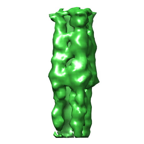
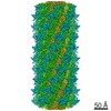
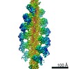
| タイトル | Mechanism of Membranous Tunnelling Nanotube Formation in Viral Genome Delivery | |||||||||
 マップデータ マップデータ | Sigma. This sub-tomogram average tube refers to the average class 3. Before displaying it, low-pass band filter to 50 Ang. | |||||||||
 試料 試料 |
| |||||||||
 キーワード キーワード | virus / structural virology / viral genome delivery / proteo-lipidic structures / membrane remodelling / nanotube formation / single-particle tomography / cellular tomography | |||||||||
| 生物種 |   Enterobacteria phage PRD1 (ファージ) Enterobacteria phage PRD1 (ファージ) | |||||||||
| 手法 | サブトモグラム平均法 / クライオ電子顕微鏡法 / 解像度: 61.0 Å | |||||||||
 データ登録者 データ登録者 | Peralta B / Gil-Carton D / Castano-Diez D / Bertin A / Boulogne C / Oksanen HM / Bamford DH / Abrescia NGA | |||||||||
 引用 引用 | ジャーナル: Nature / 年: 2004 タイトル: Membrane structure and interactions with protein and DNA in bacteriophage PRD1. 著者: Joseph J B Cockburn / Nicola G A Abrescia / Jonathan M Grimes / Geoffrey C Sutton / Jonathan M Diprose / James M Benevides / George J Thomas / Jaana K H Bamford / Dennis H Bamford / David I Stuart /  要旨: Membranes are essential for selectively controlling the passage of molecules in and out of cells and mediating the response of cells to their environment. Biological membranes and their associated ...Membranes are essential for selectively controlling the passage of molecules in and out of cells and mediating the response of cells to their environment. Biological membranes and their associated proteins present considerable difficulties for structural analysis. Although enveloped viruses have been imaged at about 9 A resolution by cryo-electron microscopy and image reconstruction, no detailed crystallographic structure of a membrane system has been described. The structure of the bacteriophage PRD1 particle, determined by X-ray crystallography at about 4 A resolution, allows the first detailed analysis of a membrane-containing virus. The architecture of the viral capsid and its implications for virus assembly are presented in the accompanying paper. Here we show that the electron density also reveals the icosahedral lipid bilayer, beneath the protein capsid, enveloping the viral DNA. The viral membrane contains about 26,000 lipid molecules asymmetrically distributed between the membrane leaflets. The inner leaflet is composed predominantly of zwitterionic phosphatidylethanolamine molecules, facilitating a very close interaction with the viral DNA, which we estimate to be packaged to a pressure of about 45 atm, factors that are likely to be important during membrane-mediated DNA translocation into the host cell. In contrast, the outer leaflet is enriched in phosphatidylglycerol and cardiolipin, which show a marked lateral segregation within the icosahedral asymmetric unit. In addition, the lipid headgroups show a surprising degree of order. | |||||||||
| 履歴 |
|
- 構造の表示
構造の表示
| ムービー |
 ムービービューア ムービービューア |
|---|---|
| 構造ビューア | EMマップ: SurfViewMolmilJmol/JSmol |
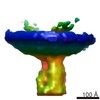
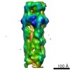
| 添付画像 |
- ダウンロードとリンク
-EMDBアーカイブ
| マップデータ | emd_2440.map.gz | 397.1 KB | EMDBマップデータ形式 | |
|---|---|---|---|---|
| ヘッダ (付随情報) | emd-2440-v30.xmlemd-2440.xml | 12.6 KB 12.6 KB | 表示 表示 | EMDBヘッダ |
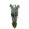
| 画像 |  EMD-2440.png EMD-2440.png | 83.8 KB | ||
| アーカイブディレクトリ |  http://ftp.pdbj.org/pub/emdb/structures/EMD-2440ftp://ftp.pdbj.org/pub/emdb/structures/EMD-2440 http://ftp.pdbj.org/pub/emdb/structures/EMD-2440ftp://ftp.pdbj.org/pub/emdb/structures/EMD-2440 | HTTPS FTP |
-検証レポート
| 文書・要旨 | emd_2440_validation.pdf.gz | 222.2 KB | 表示 | EMDB検証レポート |
|---|---|---|---|---|
| 文書・詳細版 | emd_2440_full_validation.pdf.gz | 221.3 KB | 表示 | |
| XML形式データ | emd_2440_validation.xml.gz | 5 KB | 表示 | |
| アーカイブディレクトリ | https://ftp.pdbj.org/pub/emdb/validation_reports/EMD-2440ftp://ftp.pdbj.org/pub/emdb/validation_reports/EMD-2440 | HTTPS FTP |
-関連構造データ

-リンク
| EMDBのページ | EMDB (EBI/PDBe) / EMDataResource |
|---|
-マップ
| ファイル | ダウンロード / ファイル: emd_2440.map.gz / 形式: CCP4 / 大きさ: 422.9 KB / タイプ: IMAGE STORED AS FLOATING POINT NUMBER (4 BYTES) | ||||||||||||||||||||||||||||||||||||||||||||||||||||||||||||||||||||
|---|---|---|---|---|---|---|---|---|---|---|---|---|---|---|---|---|---|---|---|---|---|---|---|---|---|---|---|---|---|---|---|---|---|---|---|---|---|---|---|---|---|---|---|---|---|---|---|---|---|---|---|---|---|---|---|---|---|---|---|---|---|---|---|---|---|---|---|---|---|
| 注釈 | Sigma. This sub-tomogram average tube refers to the average class 3. Before displaying it, low-pass band filter to 50 Ang. | ||||||||||||||||||||||||||||||||||||||||||||||||||||||||||||||||||||
| 投影像・断面図 | 画像のコントロール
画像は Spider により作成 | ||||||||||||||||||||||||||||||||||||||||||||||||||||||||||||||||||||
| ボクセルのサイズ | X=Y=Z: 8.8 Å | ||||||||||||||||||||||||||||||||||||||||||||||||||||||||||||||||||||
| 密度 |
| ||||||||||||||||||||||||||||||||||||||||||||||||||||||||||||||||||||
| 対称性 | 空間群: 1 | ||||||||||||||||||||||||||||||||||||||||||||||||||||||||||||||||||||
| 詳細 | EMDB XML:
CCP4マップ ヘッダ情報:
| ||||||||||||||||||||||||||||||||||||||||||||||||||||||||||||||||||||
 Z (Sec.)
 Y (Row.)
 X (Col.)


-添付データ
- 試料の構成要素
-全体 : Protruding tube (average class 3) from the lipid-containing bacte...
| 全体 | 名称: Protruding tube (average class 3) from the lipid-containing bacteriophage PRD1 |
|---|---|
| 要素 |
|
-超分子 #1000: Protruding tube (average class 3) from the lipid-containing bacte...
| 超分子 | 名称: Protruding tube (average class 3) from the lipid-containing bacteriophage PRD1 タイプ: sample / ID: 1000 / Number unique components: 1 |
|---|
-超分子 #1: Enterobacteria phage PRD1
| 超分子 | 名称: Enterobacteria phage PRD1 / タイプ: virus / ID: 1 / Name.synonym: bacteriophage PRD1 詳細: This is the self-assembling proteo-lipid tube. Infects both Escherichia coli and Salmonella enterica NCBI-ID: 10658 / 生物種: Enterobacteria phage PRD1 / ウイルスタイプ: VIRION / ウイルス・単離状態: SPECIES / ウイルス・エンベロープ: No / ウイルス・中空状態: No / Syn species name: bacteriophage PRD1 |
|---|---|
| 宿主 | 生物種:  |
| 分子量 | 理論値: 70 MDa |
| ウイルス殻 | Shell ID: 1 / 名称: P3 |
-実験情報
-構造解析
| 手法 | クライオ電子顕微鏡法 |
|---|---|
 解析 解析 | サブトモグラム平均法 |
| 試料の集合状態 | particle |
-試料調製
| 濃度 | 0.6 mg/mL |
|---|---|
| 緩衝液 | pH: 7.2 / 詳細: 20 mM Phosphate Buffer 1 mM MgCl2 |
| グリッド | 詳細: 200 mesh QUANTIFOIL R 2/1 (or R 3.5/1) copper grid, glow discharged in air atmosphere |
| 凍結 | 凍結剤: ETHANE / チャンバー内湿度: 95 % / チャンバー内温度: 120 K / 装置: FEI VITROBOT MARK III / 手法: Blot for 4 seconds before plunging |
- 電子顕微鏡法
| 顕微鏡 | JEOL 2200FS |
|---|---|
| 温度 | 最低: 80 K / 最高: 105 K / 平均: 99 K |
| アライメント法 | Legacy - 非点収差: Objective lens astigmatism was corrected at 120,000 times magnification |
| 特殊光学系 | エネルギーフィルター - 名称: In-column Omega filter エネルギーフィルター - エネルギー下限: 10.0 eV エネルギーフィルター - エネルギー上限: 30.0 eV |
| 日付 | 2012年1月4日 |
| 撮影 | カテゴリ: FILM フィルム・検出器のモデル: GATAN ULTRASCAN 4000 (4k x 4k) デジタル化 - スキャナー: OTHER / 平均電子線量: 100 e/Å2 / ビット/ピクセル: 16 |
| 電子線 | 加速電圧: 200 kV / 電子線源:  FIELD EMISSION GUN FIELD EMISSION GUN |
| 電子光学系 | 倍率(補正後): 34138 / 照射モード: FLOOD BEAM / 撮影モード: BRIGHT FIELD / Cs: 2 mm / 最大 デフォーカス(公称値): 8.0 µm / 最小 デフォーカス(公称値): 5.0 µm / 倍率(公称値): 25000 |
| 試料ステージ | 試料ホルダーモデル: GATAN LIQUID NITROGEN / Tilt series - Axis1 - Min angle: -64 ° / Tilt series - Axis1 - Max angle: 64 ° |
-画像解析
| 詳細 | This class 3 is one of the 4 classes used during multi-reference alignment. Please see details in primary reference. |
|---|---|
| 最終 再構成 | 想定した対称性 - 点群: C1 (非対称) / アルゴリズム: OTHER / 解像度のタイプ: BY AUTHOR / 解像度: 61.0 Å / 解像度の算出法: FSC 0.5 CUT-OFF / ソフトウェア - 名称: IMOD, Dynamo / 使用したサブトモグラム数: 64 |
