Movie
Movie Controller

[English] 日本語
 Yorodumi
- EMDB-2438: Mechanism of Membranous Tunnelling Nanotube Formation in Viral Ge... -
+ Open data

- Basic information
| Entry | Database: EMDB / ID: EMD-2438 | |||||||||
|---|---|---|---|---|---|---|---|---|---|---|
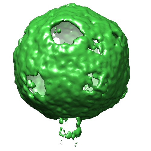
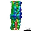
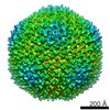
| Title | Mechanism of Membranous Tunnelling Nanotube Formation in Viral Genome Delivery | |||||||||
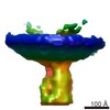
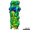
 Map data Map data | Contour level in sigma as defined by the threshold level displayed by Chimera over the RMS of the map. The recommended contour level for the visualization of the internal vesicle is about 0.35 sigma | |||||||||
 Sample Sample |
| |||||||||
 Keywords Keywords | virus / structural virology / viral genome delivery / proteo-lipidic structures / membrane remodelling / nanotube formation / single-particle tomography / cellular tomography | |||||||||
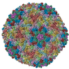
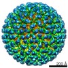
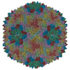
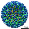
| Biological species |   Enterobacteria phage PRD1 (virus) Enterobacteria phage PRD1 (virus) | |||||||||
| Method | subtomogram averaging / cryo EM / Resolution: 64.0 Å | |||||||||
 Authors Authors | Peralta B / Gil-Carton D / Castano-Diez D / Bertin A / Boulogne C / Oksanen HM / Bamford DH / Abrescia NGA | |||||||||
 Citation Citation | Journal: Nature / Year: 2004 Title: Membrane structure and interactions with protein and DNA in bacteriophage PRD1. Authors: Joseph J B Cockburn / Nicola G A Abrescia / Jonathan M Grimes / Geoffrey C Sutton / Jonathan M Diprose / James M Benevides / George J Thomas / Jaana K H Bamford / Dennis H Bamford / David I Stuart /  Abstract: Membranes are essential for selectively controlling the passage of molecules in and out of cells and mediating the response of cells to their environment. Biological membranes and their associated ...Membranes are essential for selectively controlling the passage of molecules in and out of cells and mediating the response of cells to their environment. Biological membranes and their associated proteins present considerable difficulties for structural analysis. Although enveloped viruses have been imaged at about 9 A resolution by cryo-electron microscopy and image reconstruction, no detailed crystallographic structure of a membrane system has been described. The structure of the bacteriophage PRD1 particle, determined by X-ray crystallography at about 4 A resolution, allows the first detailed analysis of a membrane-containing virus. The architecture of the viral capsid and its implications for virus assembly are presented in the accompanying paper. Here we show that the electron density also reveals the icosahedral lipid bilayer, beneath the protein capsid, enveloping the viral DNA. The viral membrane contains about 26,000 lipid molecules asymmetrically distributed between the membrane leaflets. The inner leaflet is composed predominantly of zwitterionic phosphatidylethanolamine molecules, facilitating a very close interaction with the viral DNA, which we estimate to be packaged to a pressure of about 45 atm, factors that are likely to be important during membrane-mediated DNA translocation into the host cell. In contrast, the outer leaflet is enriched in phosphatidylglycerol and cardiolipin, which show a marked lateral segregation within the icosahedral asymmetric unit. In addition, the lipid headgroups show a surprising degree of order. | |||||||||
| History |
|
- Structure visualization
| Movie |
 Movie viewer Movie viewer |
|---|---|
| Structure viewer | EM map: SurfViewMolmilJmol/JSmol |
| Supplemental images |
- Downloads & links
-EMDB archive
| Map data | emd_2438.map.gz | 3.1 MB | EMDB map data format | |
|---|---|---|---|---|
| Header (meta data) | emd-2438-v30.xmlemd-2438.xml | 13.5 KB 13.5 KB | Display Display | EMDB header |
| Images |  EMD-2438.png EMD-2438.png | 146.6 KB | ||
| Archive directory |  http://ftp.pdbj.org/pub/emdb/structures/EMD-2438ftp://ftp.pdbj.org/pub/emdb/structures/EMD-2438 http://ftp.pdbj.org/pub/emdb/structures/EMD-2438ftp://ftp.pdbj.org/pub/emdb/structures/EMD-2438 | HTTPS FTP |
-Related structure data


-Links
| EMDB pages | EMDB (EBI/PDBe) / EMDataResource |
|---|
-Map
| File | Download / File: emd_2438.map.gz / Format: CCP4 / Size: 3.3 MB / Type: IMAGE STORED AS FLOATING POINT NUMBER (4 BYTES) | ||||||||||||||||||||||||||||||||||||||||||||||||||||||||||||||||||||
|---|---|---|---|---|---|---|---|---|---|---|---|---|---|---|---|---|---|---|---|---|---|---|---|---|---|---|---|---|---|---|---|---|---|---|---|---|---|---|---|---|---|---|---|---|---|---|---|---|---|---|---|---|---|---|---|---|---|---|---|---|---|---|---|---|---|---|---|---|---|
| Annotation | Contour level in sigma as defined by the threshold level displayed by Chimera over the RMS of the map. The recommended contour level for the visualization of the internal vesicle is about 0.35 sigma | ||||||||||||||||||||||||||||||||||||||||||||||||||||||||||||||||||||
| Projections & slices | Image control
Images are generated by Spider. | ||||||||||||||||||||||||||||||||||||||||||||||||||||||||||||||||||||
| Voxel size | X=Y=Z: 8.8 Å | ||||||||||||||||||||||||||||||||||||||||||||||||||||||||||||||||||||
| Density |
| ||||||||||||||||||||||||||||||||||||||||||||||||||||||||||||||||||||
| Symmetry | Space group: 1 | ||||||||||||||||||||||||||||||||||||||||||||||||||||||||||||||||||||
| Details | EMDB XML:
CCP4 map header:
| ||||||||||||||||||||||||||||||||||||||||||||||||||||||||||||||||||||
 Z (Sec.)
 Y (Row.)
 X (Col.)


-Supplemental data
- Sample components
-Entire : Lipid-containing bacteriophage PRD1
| Entire | Name: Lipid-containing bacteriophage PRD1 |
|---|---|
| Components |
|
-Supramolecule #1000: Lipid-containing bacteriophage PRD1
| Supramolecule | Name: Lipid-containing bacteriophage PRD1 / type: sample / ID: 1000 Details: Please keep in mind that these are PRD1 particles with a protruding tube. Therefore the icosahedral symmetry is broken and never applied during data processing and averaging Oligomeric state: A pseudo T=25 assembly / Number unique components: 1 |
|---|---|
| Molecular weight | Theoretical: 70 MDa |
-Supramolecule #1: Enterobacteria phage PRD1
| Supramolecule | Name: Enterobacteria phage PRD1 / type: virus / ID: 1 / Name.synonym: bacteriophage PRD1 Details: Infects both Escherichia coli and Salmonella enterica NCBI-ID: 10658 / Sci species name: Enterobacteria phage PRD1 / Virus type: VIRION / Virus isolate: SPECIES / Virus enveloped: No / Virus empty: No / Syn species name: bacteriophage PRD1 |
|---|---|
| Host (natural) | Organism:  |
| Molecular weight | Theoretical: 70 MDa |
| Virus shell | Shell ID: 1 / Name: P3 |
-Experimental details
-Structure determination
| Method | cryo EM |
|---|---|
 Processing Processing | subtomogram averaging |
| Aggregation state | particle |
-Sample preparation
| Concentration | 0.6 mg/mL |
|---|---|
| Buffer | pH: 7.2 / Details: 20 mM Phosphate Buffer 1 mM MgCl2 |
| Grid | Details: 200 mesh QUANTIFOIL R 2/1 (or R 3.5/1) copper grid, glow discharged in air atmosphere |
| Vitrification | Cryogen name: ETHANE / Chamber humidity: 95 % / Chamber temperature: 120 K / Instrument: FEI VITROBOT MARK III / Method: Blot for 4 seconds before plunging |
- Electron microscopy
| Microscope | JEOL 2200FS |
|---|---|
| Temperature | Min: 80 K / Max: 105 K / Average: 99 K |
| Alignment procedure | Legacy - Astigmatism: Objective lens astigmatism was corrected at 120,000 times magnification |
| Specialist optics | Energy filter - Name: In-column Omega filter / Energy filter - Lower energy threshold: 10.0 eV / Energy filter - Upper energy threshold: 30.0 eV |
| Date | Jan 4, 2012 |
| Image recording | Category: FILM / Film or detector model: GATAN ULTRASCAN 4000 (4k x 4k) / Digitization - Scanner: OTHER / Average electron dose: 100 e/Å2 / Bits/pixel: 16 |
| Electron beam | Acceleration voltage: 200 kV / Electron source:  FIELD EMISSION GUN FIELD EMISSION GUN |
| Electron optics | Calibrated magnification: 34138 / Illumination mode: FLOOD BEAM / Imaging mode: BRIGHT FIELD / Cs: 2 mm / Nominal defocus max: 8.0 µm / Nominal defocus min: 5.0 µm / Nominal magnification: 25000 |
| Sample stage | Specimen holder model: GATAN LIQUID NITROGEN / Tilt series - Axis1 - Min angle: -64 ° / Tilt series - Axis1 - Max angle: 64 ° |
-Image processing
| Details | The tube was use as pivot for initial alignment. Please see details in primary reference. |
|---|---|
| Final reconstruction | Applied symmetry - Point group: C1 (asymmetric) / Algorithm: OTHER / Resolution.type: BY AUTHOR / Resolution: 64.0 Å / Resolution method: FSC 0.5 CUT-OFF / Software - Name: IMOD, Dynamo / Number subtomograms used: 174 |
-Atomic model buiding 1
| Initial model | PDB ID: |
|---|---|
| Software | Name: Chimera |
| Details | For the docking, please, see the protocol described in the primary reference |
| Refinement | Space: REAL / Protocol: RIGID BODY FIT |