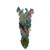
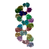
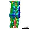
Protein or peptide: Unconventional myosin heavy chain
Ligand: ADENOSINE-5'-DIPHOSPHATE
Ligand: MAGNESIUM ION
Keywords
myosin / actin / cytoskeleton / motor protein / CONTRACTILE PROTEIN
Function / homology
Function and homology information
Regulation of CDH1 Function / Striated Muscle Contraction / actin filament-based movement / myosin complex / microfilament motor activity / striated muscle thin filament / skeletal muscle thin filament assembly / skeletal muscle fiber development / stress fiber / actin filament organization ...Regulation of CDH1 Function / Striated Muscle Contraction / actin filament-based movement / myosin complex / microfilament motor activity / striated muscle thin filament / skeletal muscle thin filament assembly / skeletal muscle fiber development / stress fiber / actin filament organization / actin filament / Hydrolases; Acting on acid anhydrides; Acting on acid anhydrides to facilitate cellular and subcellular movement / actin filament binding / actin cytoskeleton / hydrolase activity / ATP binding / membrane / cytoplasm Similarity search - Function
Plant myosin, class XI, motor domain / Class XI myosin, cargo binding domain / Dilute domain / DIL domain / Dilute domain profile. / DIL / IQ calmodulin-binding motif / Short calmodulin-binding motif containing conserved Ile and Gln residues. / IQ motif, EF-hand binding site / Myosin motor domain profile. ...Plant myosin, class XI, motor domain / Class XI myosin, cargo binding domain / Dilute domain / DIL domain / Dilute domain profile. / DIL / IQ calmodulin-binding motif / Short calmodulin-binding motif containing conserved Ile and Gln residues. / IQ motif, EF-hand binding site / Myosin motor domain profile. / Myosin head, motor domain / Myosin head (motor domain) / Myosin. Large ATPases. / IQ motif profile. / Kinesin motor domain superfamily / Actins signature 1. / Actin, conserved site / Actins signature 2. / Actin/actin-like conserved site / Actins and actin-related proteins signature. / Actin / Actin family / Actin / ATPase, nucleotide binding domain / P-loop containing nucleoside triphosphate hydrolase Similarity search - Domain/homology
National Institutes of Health/National Institute of General Medical Sciences (NIH/NIGMS)
R01-GM114627
United States
Citation
Journal: Nat Chem Biol / Year: 2021 Title: Optical control of fast and processive engineered myosins in vitro and in living cells. Authors: Paul V Ruijgrok / Rajarshi P Ghosh / Sasha Zemsky / Muneaki Nakamura / Rui Gong / Lin Ning / Robert Chen / Vipul T Vachharajani / Alexander E Chu / Namrata Anand / Raphael R Eguchi / Po-Ssu ...Authors: Paul V Ruijgrok / Rajarshi P Ghosh / Sasha Zemsky / Muneaki Nakamura / Rui Gong / Lin Ning / Robert Chen / Vipul T Vachharajani / Alexander E Chu / Namrata Anand / Raphael R Eguchi / Po-Ssu Huang / Michael Z Lin / Gregory M Alushin / Jan T Liphardt / Zev Bryant / Abstract: Precision tools for spatiotemporal control of cytoskeletal motor function are needed to dissect fundamental biological processes ranging from intracellular transport to cell migration and division. ...Precision tools for spatiotemporal control of cytoskeletal motor function are needed to dissect fundamental biological processes ranging from intracellular transport to cell migration and division. Direct optical control of motor speed and direction is one promising approach, but it remains a challenge to engineer controllable motors with desirable properties such as the speed and processivity required for transport applications in living cells. Here, we develop engineered myosin motors that combine large optical modulation depths with high velocities, and create processive myosin motors with optically controllable directionality. We characterize the performance of the motors using in vitro motility assays, single-molecule tracking and live-cell imaging. Bidirectional processive motors move efficiently toward the tips of cellular protrusions in the presence of blue light, and can transport molecular cargo in cells. Robust gearshifting myosins will further enable programmable transport in contexts ranging from in vitro active matter reconstitutions to microfabricated systems that harness molecular propulsion.
History
Deposition
Oct 5, 2020
-
Header (metadata) release
Jan 13, 2021
-
Map release
Jan 13, 2021
-
Update
Mar 6, 2024
-
Current status
Mar 6, 2024
Processing site: RCSB / Status: Released
-
Structure visualization
Movie
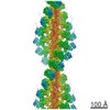
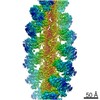
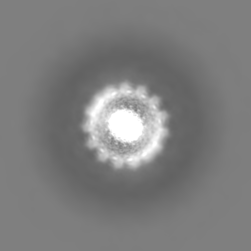
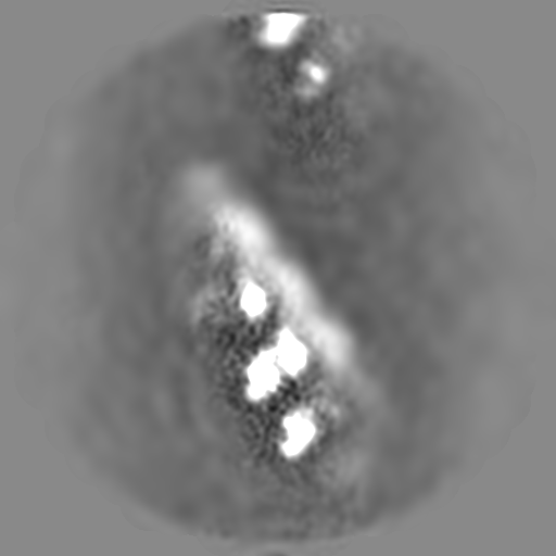
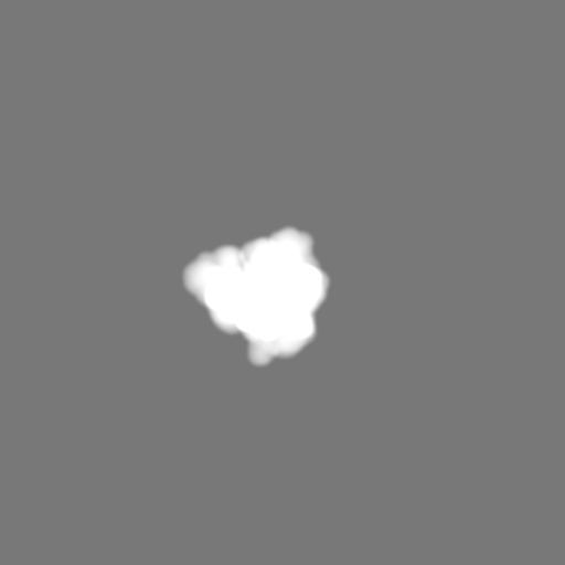
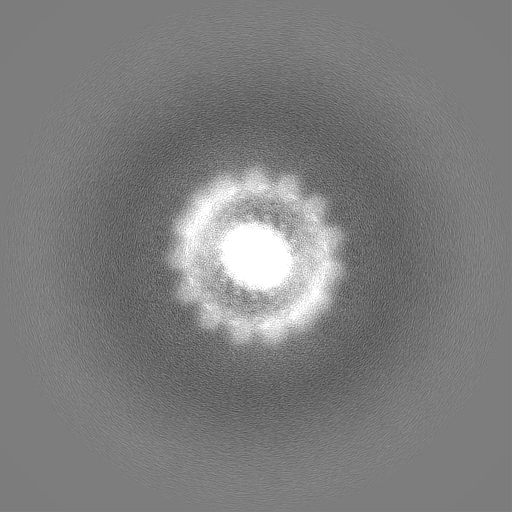
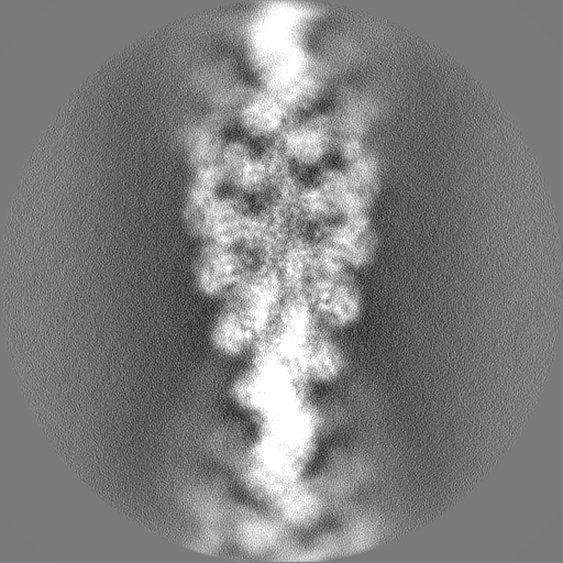
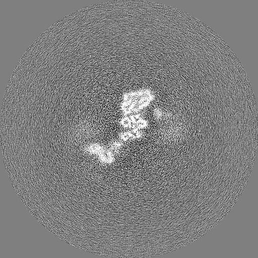
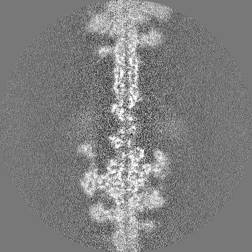
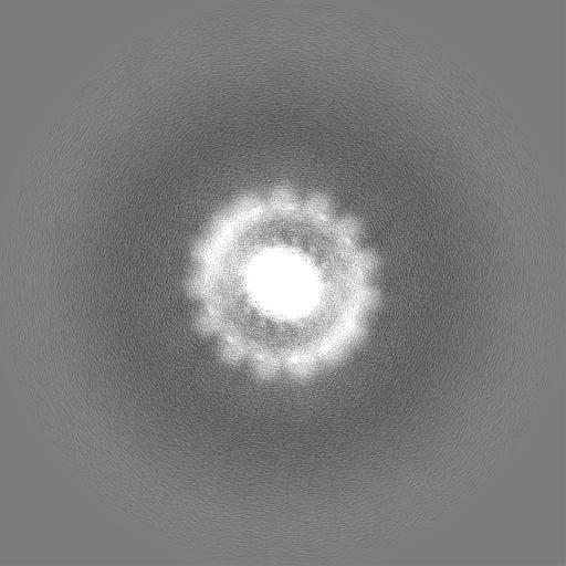
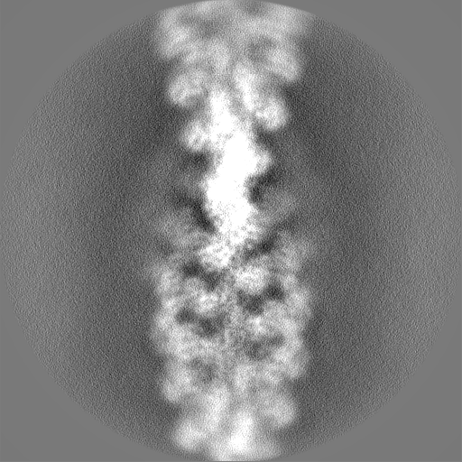
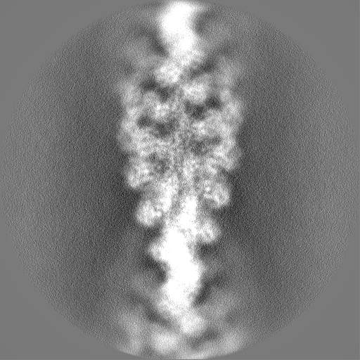
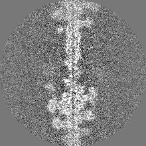
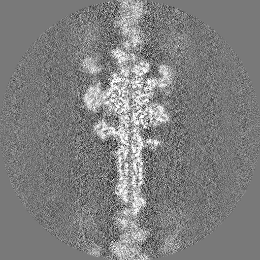
Surface view with section colored by density value
Name: MAGNESIUM ION / type: ligand / ID: 4 / Number of copies: 3 / Formula: MG
Molecular weight
Theoretical: 24.305 Da
-
Experimental details
-
Structure determination
Method
cryo EM
Processing
helical reconstruction
Aggregation state
filament
-
Sample preparation
Buffer
pH: 7 Component:
Concentration
Formula
Name
10.0 mM
C3N2H4
Imidazole
50.0 mM
KCl
Potassium chloride
1.0 mM
MgCl2
Magnesium chloride
1.0 mM
C14H24N2O10
EGTA
0.5 mM
C4H10O2S2
DTT
0.01 weight/volume
NaN3
sodium azide
Details: 10 mM imidazole pH 7.0,50 mM KCl,1mM MgCl2, 1mM EGTA, 0.5 mM DTT, 0.01% NaN3
Grid
Model: C-flat-1.2/1.3 / Material: GOLD / Mesh: 300 / Support film - Material: CARBON / Support film - topology: HOLEY / Support film - Film thickness: 20 / Pretreatment - Type: PLASMA CLEANING / Pretreatment - Time: 10 sec. / Pretreatment - Atmosphere: OTHER
Vitrification
Cryogen name: ETHANE / Chamber humidity: 100 % / Chamber temperature: 293 K / Instrument: LEICA EM GP
-
Electron microscopy
Microscope
FEI TITAN KRIOS
Image recording
Film or detector model: GATAN K2 SUMMIT (4k x 4k) / Detector mode: COUNTING / Average electron dose: 67.12 e/Å2
Electron beam
Acceleration voltage: 300 kV / Electron source: FIELD EMISSION GUN
In the structure databanks used in Yorodumi, some data are registered as the other names, "COVID-19 virus" and "2019-nCoV". Here are the details of the virus and the list of structure data.
Jan 31, 2019. EMDB accession codes are about to change! (news from PDBe EMDB page)
EMDB accession codes are about to change! (news from PDBe EMDB page)
The allocation of 4 digits for EMDB accession codes will soon come to an end. Whilst these codes will remain in use, new EMDB accession codes will include an additional digit and will expand incrementally as the available range of codes is exhausted. The current 4-digit format prefixed with “EMD-” (i.e. EMD-XXXX) will advance to a 5-digit format (i.e. EMD-XXXXX), and so on. It is currently estimated that the 4-digit codes will be depleted around Spring 2019, at which point the 5-digit format will come into force.
The EM Navigator/Yorodumi systems omit the EMD- prefix.
Related info.:Q: What is EMD? / ID/Accession-code notation in Yorodumi/EM Navigator
Yorodumi is a browser for structure data from EMDB, PDB, SASBDB, etc.
This page is also the successor to EM Navigator detail page, and also detail information page/front-end page for Omokage search.
The word "yorodu" (or yorozu) is an old Japanese word meaning "ten thousand". "mi" (miru) is to see.
Related info.:EMDB / PDB / SASBDB / Comparison of 3 databanks / Yorodumi Search / Aug 31, 2016. New EM Navigator & Yorodumi / Yorodumi Papers / Jmol/JSmol / Function and homology information / Changes in new EM Navigator and Yorodumi
 Movie
Movie Controller


 Open data
 Basic information
 Map data
 Sample
 Keywords
 Function and homology information
 Chara corallina (plant)
 Authors
 United States, 1 items
  Citation
 Structure visualization
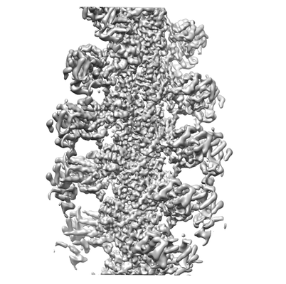
 Downloads & links
 emd_22808.png
 http://ftp.pdbj.org/pub/emdb/structures/EMD-22808


 Z (Sec.)
 Y (Row.)
 X (Col.)


 Sample components
 Spodoptera aff. frugiperda 2 RZ-2014 (butterflies/moths)
 Processing
 Electron microscopy
 FIELD EMISSION GUN