 Movie
Movie Controller


+ Open data

- Basic information
| Entry | Database: EMDB / ID: EMD-2433 | |||||||||
|---|---|---|---|---|---|---|---|---|---|---|
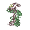
| Title | Amyloid-beta nanotube | |||||||||
 Map data Map data | Reconstruction of an amyloid-beta nanotube | |||||||||
 Sample Sample |
| |||||||||
 Keywords Keywords | nanotube / neurodegeneration / prion-dependent synaptotoxicity | |||||||||
| Biological species |  Homo sapiens (human) Homo sapiens (human) | |||||||||
| Method | single particle reconstruction / negative staining | |||||||||
 Authors Authors | Nicoll AJ / Panico S / Freir DB / Wright D / Terry C / Risse E / Herron CE / O'Malley T / Wadsworth JD / Farrow MA ...Nicoll AJ / Panico S / Freir DB / Wright D / Terry C / Risse E / Herron CE / O'Malley T / Wadsworth JD / Farrow MA / Walsh DM / Saibil HR / Collinge J | |||||||||
 Citation Citation | Journal: Nat Commun / Year: 2013 Title: Amyloid-β nanotubes are associated with prion protein-dependent synaptotoxicity. Authors: Andrew J Nicoll / Silvia Panico / Darragh B Freir / Daniel Wright / Cassandra Terry / Emmanuel Risse / Caroline E Herron / Tiernan O'Malley / Jonathan D F Wadsworth / Mark A Farrow / Dominic ...Authors: Andrew J Nicoll / Silvia Panico / Darragh B Freir / Daniel Wright / Cassandra Terry / Emmanuel Risse / Caroline E Herron / Tiernan O'Malley / Jonathan D F Wadsworth / Mark A Farrow / Dominic M Walsh / Helen R Saibil / John Collinge /  Abstract: Growing evidence suggests water-soluble, non-fibrillar forms of amyloid-β protein (Aβ) have important roles in Alzheimer's disease with toxicities mimicked by synthetic Aβ(1-42). However, no ...Growing evidence suggests water-soluble, non-fibrillar forms of amyloid-β protein (Aβ) have important roles in Alzheimer's disease with toxicities mimicked by synthetic Aβ(1-42). However, no defined toxic structures acting via specific receptors have been identified and roles of proposed receptors, such as prion protein (PrP), remain controversial. Here we quantify binding to PrP of Aβ(1-42) after different durations of aggregation. We show PrP-binding and PrP-dependent inhibition of long-term potentiation (LTP) correlate with the presence of protofibrils. Globular oligomers bind less avidly to PrP and do not inhibit LTP, whereas fibrils inhibit LTP in a PrP-independent manner. That only certain transient Aβ assemblies cause PrP-dependent toxicity explains conflicting reports regarding the involvement of PrP in Aβ-induced impairments. We show that these protofibrils contain a defined nanotubular structure with a previously unidentified triple helical conformation. Blocking the formation of Aβ nanotubes or their interaction with PrP might have a role in treatment of Alzheimer's disease. | |||||||||
| History |
|
- Structure visualization
| Movie |
 Movie viewer Movie viewer |
|---|---|
| Structure viewer | EM map: SurfViewMolmilJmol/JSmol |
| Supplemental images |
 UCSF Chimera
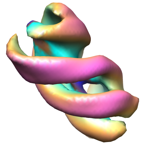
- Downloads & links
-EMDB archive
| Map data | emd_2433.map.gz | 241.1 KB | EMDB map data format | |
|---|---|---|---|---|
| Header (meta data) | emd-2433-v30.xmlemd-2433.xml | 10.9 KB 10.9 KB | Display Display | EMDB header |
| Images |  emd_2433.png emd_2433.png | 116.6 KB | ||
| Archive directory |  http://ftp.pdbj.org/pub/emdb/structures/EMD-2433ftp://ftp.pdbj.org/pub/emdb/structures/EMD-2433 http://ftp.pdbj.org/pub/emdb/structures/EMD-2433ftp://ftp.pdbj.org/pub/emdb/structures/EMD-2433 | HTTPS FTP |
-Validation report
| Summary document | emd_2433_validation.pdf.gz | 195.2 KB | Display | EMDB validaton report |
|---|---|---|---|---|
| Full document | emd_2433_full_validation.pdf.gz | 194.3 KB | Display | |
| Data in XML | emd_2433_validation.xml.gz | 4.2 KB | Display | |
| Arichive directory | https://ftp.pdbj.org/pub/emdb/validation_reports/EMD-2433ftp://ftp.pdbj.org/pub/emdb/validation_reports/EMD-2433 | HTTPS FTP |
-Related structure data
| Similar structure data |
|---|
-Links
| EMDB pages | EMDB (EBI/PDBe) / EMDataResource |
|---|---|
| Related items in Molecule of the Month |

-Map
| File | Download / File: emd_2433.map.gz / Format: CCP4 / Size: 459 KB / Type: IMAGE STORED AS FLOATING POINT NUMBER (4 BYTES) | ||||||||||||||||||||||||||||||||||||||||||||||||||||||||||||||||||||
|---|---|---|---|---|---|---|---|---|---|---|---|---|---|---|---|---|---|---|---|---|---|---|---|---|---|---|---|---|---|---|---|---|---|---|---|---|---|---|---|---|---|---|---|---|---|---|---|---|---|---|---|---|---|---|---|---|---|---|---|---|---|---|---|---|---|---|---|---|---|
| Annotation | Reconstruction of an amyloid-beta nanotube | ||||||||||||||||||||||||||||||||||||||||||||||||||||||||||||||||||||
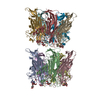
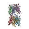
| Projections & slices | Image control
Images are generated by Spider. generated in cubic-lattice coordinate | ||||||||||||||||||||||||||||||||||||||||||||||||||||||||||||||||||||
| Voxel size | X=Y=Z: 3.26 Å | ||||||||||||||||||||||||||||||||||||||||||||||||||||||||||||||||||||
| Density |
| ||||||||||||||||||||||||||||||||||||||||||||||||||||||||||||||||||||
| Symmetry | Space group: 1 | ||||||||||||||||||||||||||||||||||||||||||||||||||||||||||||||||||||
| Details | EMDB XML:
CCP4 map header:
| ||||||||||||||||||||||||||||||||||||||||||||||||||||||||||||||||||||
 Z (Sec.)
 Y (Row.)
 X (Col.)


-Supplemental data
- Sample components
-Entire : Amyloid-Beta (1-42) nanotube
| Entire | Name: Amyloid-Beta (1-42) nanotube |
|---|---|
| Components |
|
-Supramolecule #1000: Amyloid-Beta (1-42) nanotube
| Supramolecule | Name: Amyloid-Beta (1-42) nanotube / type: sample / ID: 1000 Details: Protofibrils were prepared from synthetic Amyloid-Beta (1-42) peptide. Briefly hexafluoro-2-propanol-trated Amyloid-beta (1-42) was dissolved in DMSO, diluted into phenol red-free Hams-F12 ...Details: Protofibrils were prepared from synthetic Amyloid-Beta (1-42) peptide. Briefly hexafluoro-2-propanol-trated Amyloid-beta (1-42) was dissolved in DMSO, diluted into phenol red-free Hams-F12 medium, centrifuged and incubated at 22 C for 16 hours. Size of protofibrils ranged from 0.1 to 1 megadaltons Oligomeric state: protofibrils / Number unique components: 1 |
|---|---|
| Molecular weight | Experimental: 500 KDa Method: size-exclusion chromatography with static light scattering |
-Macromolecule #1: Amyloid-beta (1-42)
| Macromolecule | Name: Amyloid-beta (1-42) / type: protein_or_peptide / ID: 1 / Recombinant expression: No / Database: NCBI |
|---|---|
| Source (natural) | Organism: Homo sapiens (human) / synonym: human |
-Experimental details
-Structure determination
| Method | negative staining |
|---|---|
 Processing Processing | single particle reconstruction |
| Aggregation state | filament |
-Sample preparation
| Concentration | 0.1 mg/mL |
|---|---|
| Buffer | Details: phenol red-free Ham's F12 cell medium (2% DMSO) |
| Staining | Type: NEGATIVE Details: Grids with adsorbed protein were stained with 2% w/v uranyl acetate for 1 minute |
| Grid | Details: Negatively glow discharged 300 mesh copper grid with continuous carbon layer |
| Vitrification | Cryogen name: NONE / Instrument: OTHER |
- Electron microscopy
| Microscope | FEI TECNAI 12 |
|---|---|
| Alignment procedure | Legacy - Astigmatism: corrected for at specimen level |
| Date | Jan 10, 2011 |
| Image recording | Category: FILM / Film or detector model: KODAK SO-163 FILM / Digitization - Scanner: ZEISS SCAI / Digitization - Sampling interval: 7 µm / Number real images: 50 / Average electron dose: 20 e/Å2 / Bits/pixel: 8 |
| Electron beam | Acceleration voltage: 120 kV / Electron source: TUNGSTEN HAIRPIN |
| Electron optics | Calibrated magnification: 42986 / Illumination mode: FLOOD BEAM / Imaging mode: BRIGHT FIELD / Cs: 2.2 mm / Nominal defocus max: 1.0 µm / Nominal defocus min: 0.7 µm / Nominal magnification: 42000 |
| Sample stage | Specimen holder model: SIDE ENTRY, EUCENTRIC |
-Image processing
| Details | Three-dimensional reconstructions were calculated from extracted, aligned and classified segments and applying helical symmetry, using the pitch independently determined from tomography and single pixel increments in z to generate continuous helices. |
|---|---|
| CTF correction | Details: phase flipping, whole micrograph |
| Final reconstruction | Applied symmetry - Helical parameters - Δz: 3.26 Å Applied symmetry - Helical parameters - Δ&Phi: 15 ° Algorithm: OTHER / Software - Name: Spider Details: Maps were calculated from individual classes. Final volume is the average of six maps (maps averaged in CHIMERA). Helical pitch repeat was obtained from tomography data. Since the subunit ...Details: Maps were calculated from individual classes. Final volume is the average of six maps (maps averaged in CHIMERA). Helical pitch repeat was obtained from tomography data. Since the subunit repeat could not be determined, continuous helices were generated. Number images used: 155 |
| Final two d classification | Number classes: 6 |
