 Movie
Movie Controller


+ Open data

- Basic information
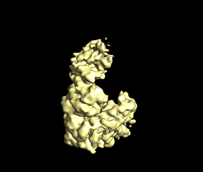
| Entry | Database: EMDB / ID: EMD-24329 | |||||||||||||||
|---|---|---|---|---|---|---|---|---|---|---|---|---|---|---|---|---|
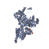
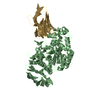
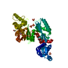
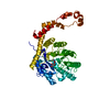
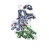
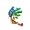
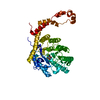
| Title | Methanococcus maripaludis chaperonin, closed conformation 2 | |||||||||||||||
 Map data Map data | ||||||||||||||||
 Sample Sample |
| |||||||||||||||
 Keywords Keywords | Open conformation / CHAPERONE | |||||||||||||||
| Function / homology |  Function and homology information Function and homology informationchaperonin ATPase / Hydrolases; Acting on acid anhydrides; In phosphorus-containing anhydrides / ATP-dependent protein folding chaperone / : / ATP hydrolysis activity / protein-containing complex / ATP binding / cytoplasm Similarity search - Function | |||||||||||||||
| Biological species |  Methanococcus maripaludis (archaea) Methanococcus maripaludis (archaea) | |||||||||||||||
| Method | single particle reconstruction / cryo EM / Resolution: 4.0 Å | |||||||||||||||
 Authors Authors | Zhao Y / Schmid M | |||||||||||||||
| Funding support |  United States, 4 items United States, 4 items
| |||||||||||||||
 Citation Citation | Journal: Nat Commun / Year: 2021 Title: CryoEM reveals the stochastic nature of individual ATP binding events in a group II chaperonin. Authors: Yanyan Zhao / Michael F Schmid / Judith Frydman / Wah Chiu / Abstract: Chaperonins are homo- or hetero-oligomeric complexes that use ATP binding and hydrolysis to facilitate protein folding. ATP hydrolysis exhibits both positive and negative cooperativity. The mechanism ...Chaperonins are homo- or hetero-oligomeric complexes that use ATP binding and hydrolysis to facilitate protein folding. ATP hydrolysis exhibits both positive and negative cooperativity. The mechanism by which chaperonins coordinate ATP utilization in their multiple subunits remains unclear. Here we use cryoEM to study ATP binding in the homo-oligomeric archaeal chaperonin from Methanococcus maripaludis (MmCpn), consisting of two stacked rings composed of eight identical subunits each. Using a series of image classification steps, we obtained different structural snapshots of individual chaperonins undergoing the nucleotide binding process. We identified nucleotide-bound and free states of individual subunits in each chaperonin, allowing us to determine the ATP occupancy state of each MmCpn particle. We observe distinctive tertiary and quaternary structures reflecting variations in nucleotide occupancy and subunit conformations in each chaperonin complex. Detailed analysis of the nucleotide distribution in each MmCpn complex indicates that individual ATP binding events occur in a statistically random manner for MmCpn, both within and across the rings. Our findings illustrate the power of cryoEM to characterize a biochemical property of multi-subunit ligand binding cooperativity at the individual particle level. | |||||||||||||||
| History |
|
- Structure visualization
| Movie |
Movie viewer |
|---|---|
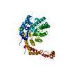
| Structure viewer | EM map: SurfViewMolmilJmol/JSmol |
| Supplemental images |
- Downloads & links
-EMDB archive
| Map data | emd_24329.map.gz | 2.5 MB | EMDB map data format | |
|---|---|---|---|---|
| Header (meta data) | emd-24329-v30.xmlemd-24329.xml | 11.5 KB 11.5 KB | Display Display | EMDB header |
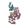
| Images |  emd_24329.png emd_24329.png | 49.1 KB | ||
| Filedesc metadata | emd-24329.cif.gz | 5.1 KB | ||
| Archive directory |  http://ftp.pdbj.org/pub/emdb/structures/EMD-24329ftp://ftp.pdbj.org/pub/emdb/structures/EMD-24329 http://ftp.pdbj.org/pub/emdb/structures/EMD-24329ftp://ftp.pdbj.org/pub/emdb/structures/EMD-24329 | HTTPS FTP |
-Related structure data
| Related structure data |  7r9mMC  7r9eC  7r9hC  7r9iC  7r9jC  7r9kC  7r9oC  7r9uC  7rakC C: citing same article ( M: atomic model generated by this map |
|---|---|
| Similar structure data | |
| EM raw data | EMPIAR-10770 (Title: Cryo-EM reveals the stochastic nature of individual ATP binding events in a group II chaperonin Data size: 48.4 Data #1: aligned particle image [picked particles - single frame - processed]) |


-Links
| EMDB pages | EMDB (EBI/PDBe) / EMDataResource |
|---|---|
| Related items in Molecule of the Month |

-Map
| File | Download / File: emd_24329.map.gz / Format: CCP4 / Size: 83.7 MB / Type: IMAGE STORED AS FLOATING POINT NUMBER (4 BYTES) | ||||||||||||||||||||||||||||||||||||||||||||||||||||||||||||||||||||
|---|---|---|---|---|---|---|---|---|---|---|---|---|---|---|---|---|---|---|---|---|---|---|---|---|---|---|---|---|---|---|---|---|---|---|---|---|---|---|---|---|---|---|---|---|---|---|---|---|---|---|---|---|---|---|---|---|---|---|---|---|---|---|---|---|---|---|---|---|---|
| Projections & slices | Image control
Images are generated by Spider. | ||||||||||||||||||||||||||||||||||||||||||||||||||||||||||||||||||||
| Voxel size | X=Y=Z: 1.08 Å | ||||||||||||||||||||||||||||||||||||||||||||||||||||||||||||||||||||
| Density |
| ||||||||||||||||||||||||||||||||||||||||||||||||||||||||||||||||||||
| Symmetry | Space group: 1 | ||||||||||||||||||||||||||||||||||||||||||||||||||||||||||||||||||||
| Details | EMDB XML:
CCP4 map header:
| ||||||||||||||||||||||||||||||||||||||||||||||||||||||||||||||||||||
 Z (Sec.)
 Y (Row.)
 X (Col.)


-Supplemental data
- Sample components
-Entire : MmCpn
| Entire | Name: MmCpn |
|---|---|
| Components |
|
-Supramolecule #1: MmCpn
| Supramolecule | Name: MmCpn / type: complex / ID: 1 / Parent: 0 / Macromolecule list: all |
|---|---|
| Source (natural) | Organism: Methanococcus maripaludis (archaea) |
| Molecular weight | Theoretical: 1 MDa |
-Macromolecule #1: Chaperonin
| Macromolecule | Name: Chaperonin / type: protein_or_peptide / ID: 1 / Number of copies: 1 / Enantiomer: LEVO |
|---|---|
| Source (natural) | Organism: Methanococcus maripaludis (archaea) |
| Molecular weight | Theoretical: 54.372539 KDa |
| Recombinant expression | Organism:  |
| Sequence | String: RDAQRMNILA GRIIAETVRS TLGPKGMDKM LVDDLGDVVV TNDGVTILRE MSVEHPAAKM LIEVAKTQEK EVGDGTTTAV VVAGELLRK AEELLDQNVH PTIVVKGYQA AAQKAQELLK TIACEVGAQD KEILTKIAMT SITGKGAEKA KEKLAEIIVE A VSAVVDDE ...String: RDAQRMNILA GRIIAETVRS TLGPKGMDKM LVDDLGDVVV TNDGVTILRE MSVEHPAAKM LIEVAKTQEK EVGDGTTTAV VVAGELLRK AEELLDQNVH PTIVVKGYQA AAQKAQELLK TIACEVGAQD KEILTKIAMT SITGKGAEKA KEKLAEIIVE A VSAVVDDE GKVDKDLIKI EKKSGASIDD TELIKGVLVD KERVSAQMPK KVTDAKIALL NCAIEIKETE TDAEIRITDP AK LMEFIEQ EEKMLKDMVA EIKASGANVL FCQKGIDDLA QHYLAKEGIV AARRVKKSDM EKLAKATGAN VITNIKDLSA QDL GDAGLV EERKISGDSM IFVEECKHPK AVTMLIRGTT EHVIEEVARA VDDAVGVVGC TIEDGRIVSG GGSTEVELSM KLRE YAEGI SGREQLAVRA FADALEVIPR TLAENAGLDA IEILVKVRAA HASNGNKCAG LNVFTGAVED MCENGVVEPL RVKTQ AIQS AAESTEMLLR IDDVIAAEKL R UniProtKB: Chaperonin subunit |
-Experimental details
-Structure determination
| Method | cryo EM |
|---|---|
 Processing Processing | single particle reconstruction |
| Aggregation state | particle |
-Sample preparation
| Buffer | pH: 7.4 |
|---|---|
| Vitrification | Cryogen name: ETHANE |
- Electron microscopy
| Microscope | FEI TITAN KRIOS |
|---|---|
| Image recording | Film or detector model: GATAN K2 SUMMIT (4k x 4k) / Detector mode: COUNTING / Average electron dose: 50.0 e/Å2 |
| Electron beam | Acceleration voltage: 300 kV / Electron source:  FIELD EMISSION GUN FIELD EMISSION GUN |
| Electron optics | Illumination mode: FLOOD BEAM / Imaging mode: BRIGHT FIELD |
| Experimental equipment |  Model: Titan Krios / Image courtesy: FEI Company |
-Image processing
| Startup model | Type of model: NONE |
|---|---|
| Final reconstruction | Resolution.type: BY AUTHOR / Resolution: 4.0 Å / Resolution method: FSC 0.143 CUT-OFF / Number images used: 233000 |
| Initial angle assignment | Type: MAXIMUM LIKELIHOOD |
| Final angle assignment | Type: MAXIMUM LIKELIHOOD |
-Atomic model buiding 1
| Refinement | Protocol: FLEXIBLE FIT |
|---|---|
| Output model | PDB-7r9m: |
