+ データを開く
- 基本情報
基本情報
| 登録情報 | データベース: PDB / ID: 7r9h | |||||||||||||||
|---|---|---|---|---|---|---|---|---|---|---|---|---|---|---|---|---|
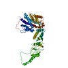
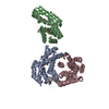
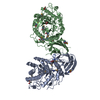
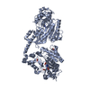
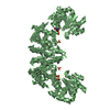
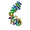
| タイトル | Methanococcus maripaludis chaperonin, open conformation 2 | |||||||||||||||
 要素 要素 | Chaperonin | |||||||||||||||
 キーワード キーワード | CHAPERONE / Open conformation | |||||||||||||||
| 機能・相同性 |  機能・相同性情報 機能・相同性情報ATP-dependent protein folding chaperone / unfolded protein binding / ATP hydrolysis activity / protein-containing complex / ATP binding / metal ion binding / identical protein binding / cytoplasm 類似検索 - 分子機能 | |||||||||||||||
| 生物種 |  Methanococcus maripaludis (古細菌) Methanococcus maripaludis (古細菌) | |||||||||||||||
| 手法 | 電子顕微鏡法 / 単粒子再構成法 / クライオ電子顕微鏡法 / 解像度: 6.3 Å | |||||||||||||||
 データ登録者 データ登録者 | Zhao, Y. / Schmid, M. / Frydman, J. / Chiu, W. | |||||||||||||||
| 資金援助 |  米国, 4件 米国, 4件
| |||||||||||||||
 引用 引用 |  ジャーナル: Nat Commun / 年: 2021 ジャーナル: Nat Commun / 年: 2021タイトル: CryoEM reveals the stochastic nature of individual ATP binding events in a group II chaperonin. 著者: Yanyan Zhao / Michael F Schmid / Judith Frydman / Wah Chiu /  要旨: Chaperonins are homo- or hetero-oligomeric complexes that use ATP binding and hydrolysis to facilitate protein folding. ATP hydrolysis exhibits both positive and negative cooperativity. The mechanism ...Chaperonins are homo- or hetero-oligomeric complexes that use ATP binding and hydrolysis to facilitate protein folding. ATP hydrolysis exhibits both positive and negative cooperativity. The mechanism by which chaperonins coordinate ATP utilization in their multiple subunits remains unclear. Here we use cryoEM to study ATP binding in the homo-oligomeric archaeal chaperonin from Methanococcus maripaludis (MmCpn), consisting of two stacked rings composed of eight identical subunits each. Using a series of image classification steps, we obtained different structural snapshots of individual chaperonins undergoing the nucleotide binding process. We identified nucleotide-bound and free states of individual subunits in each chaperonin, allowing us to determine the ATP occupancy state of each MmCpn particle. We observe distinctive tertiary and quaternary structures reflecting variations in nucleotide occupancy and subunit conformations in each chaperonin complex. Detailed analysis of the nucleotide distribution in each MmCpn complex indicates that individual ATP binding events occur in a statistically random manner for MmCpn, both within and across the rings. Our findings illustrate the power of cryoEM to characterize a biochemical property of multi-subunit ligand binding cooperativity at the individual particle level. | |||||||||||||||
| 履歴 |
|
- 構造の表示
構造の表示
| ムービー |
 ムービービューア ムービービューア |
|---|---|
| 構造ビューア | 分子:  Molmil Molmil Jmol/JSmol Jmol/JSmol |
- ダウンロードとリンク
- ダウンロード
| PDBx/mmCIF形式 |  7r9h.cif.gz 7r9h.cif.gz | 168.3 KB | 表示 |  PDBx/mmCIF形式 PDBx/mmCIF形式 |
|---|---|---|---|---|
| PDB形式 |  pdb7r9h.ent.gz pdb7r9h.ent.gz | 131.6 KB | 表示 |  PDB形式 PDB形式 |
| PDBx/mmJSON形式 |  7r9h.json.gz 7r9h.json.gz | ツリー表示 |  PDBx/mmJSON形式 PDBx/mmJSON形式 | |
| その他 |  その他のダウンロード その他のダウンロード |
-検証レポート
| アーカイブディレクトリ |  https://data.pdbj.org/pub/pdb/validation_reports/r9/7r9h https://data.pdbj.org/pub/pdb/validation_reports/r9/7r9h ftp://data.pdbj.org/pub/pdb/validation_reports/r9/7r9h ftp://data.pdbj.org/pub/pdb/validation_reports/r9/7r9h | HTTPS FTP |
|---|
-関連構造データ
| 関連構造データ |  24325MC  7r9eC  7r9iC  7r9jC  7r9kC  7r9mC  7r9oC  7r9uC  7rakC C: 同じ文献を引用 ( M: このデータのモデリングに利用したマップデータ |
|---|---|
| 類似構造データ | |
| 電子顕微鏡画像生データ |  EMPIAR-10770 (タイトル: Cryo-EM reveals the stochastic nature of individual ATP binding events in a group II chaperonin EMPIAR-10770 (タイトル: Cryo-EM reveals the stochastic nature of individual ATP binding events in a group II chaperoninData size: 48.4 Data #1: aligned particle image [picked particles - single frame - processed]) |
- リンク
リンク
- 集合体
集合体
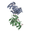
| 登録構造単位 | 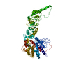
|
|---|---|
| 1 |
|
- 要素
要素
| #1: タンパク質 | 分子量: 54372.539 Da / 分子数: 1 / 由来タイプ: 組換発現 由来: (組換発現)  Methanococcus maripaludis (古細菌) Methanococcus maripaludis (古細菌)発現宿主:  |
|---|
-実験情報
-実験
| 実験 | 手法: 電子顕微鏡法 |
|---|---|
| EM実験 | 試料の集合状態: PARTICLE / 3次元再構成法: 単粒子再構成法 |
- 試料調製
試料調製
| 構成要素 | 名称: MmCpn / タイプ: COMPLEX / Entity ID: all / 由来: RECOMBINANT |
|---|---|
| 分子量 | 値: 1 MDa |
| 由来(天然) | 生物種:  Methanococcus maripaludis (古細菌) Methanococcus maripaludis (古細菌) |
| 由来(組換発現) | 生物種:  |
| 緩衝液 | pH: 7.4 |
| 試料 | 包埋: NO / シャドウイング: NO / 染色: NO / 凍結: YES |
| 急速凍結 | 凍結剤: ETHANE |
- 電子顕微鏡撮影
| 実験機器 |  モデル: Titan Krios / 画像提供: FEI Company |
|---|---|
| 顕微鏡 | モデル: FEI TITAN KRIOS |
| 電子銃 | 電子線源:  FIELD EMISSION GUN / 加速電圧: 300 kV / 照射モード: FLOOD BEAM FIELD EMISSION GUN / 加速電圧: 300 kV / 照射モード: FLOOD BEAM |
| 電子レンズ | モード: BRIGHT FIELD |
| 撮影 | 電子線照射量: 50 e/Å2 / 検出モード: COUNTING フィルム・検出器のモデル: GATAN K2 SUMMIT (4k x 4k) |
- 解析
解析
| CTF補正 | タイプ: PHASE FLIPPING ONLY |
|---|---|
| 3次元再構成 | 解像度: 6.3 Å / 解像度の算出法: FSC 0.143 CUT-OFF / 粒子像の数: 3800 / 対称性のタイプ: POINT |
| 原子モデル構築 | プロトコル: FLEXIBLE FIT |
 ムービー
ムービー コントローラー


 PDBj
PDBj
