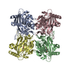
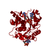
- PDB-1z05: Crystal structure of the ROK family transcriptional regulator, ho... -
+
Open data
ID or keywords:
Loading...
-
Basic information
Entry
Database: PDB / ID: 1z05
Title
Crystal structure of the ROK family transcriptional regulator, homolog of E.coli MLC protein.
Components
transcriptional regulator, ROK family
Keywords
TRANSCRIPTION / ROK family / transcriptional regulator / Structural Genomics / PSI / Protein Structure Initiative / Midwest Center for Structural Genomics / MCSG
Function / homology
Function and homology information
regulation of DNA-templated transcription / DNA-templated transcription / DNA binding / metal ion binding Similarity search - Function
: / ROK family signature. / ROK family / ROK family / ATPase, nucleotide binding domain / Winged helix-like DNA-binding domain superfamily/Winged helix DNA-binding domain / ATPase, nucleotide binding domain / Nucleotidyltransferase; domain 5 / Arc Repressor Mutant, subunit A / Winged helix DNA-binding domain superfamily ...: / ROK family signature. / ROK family / ROK family / ATPase, nucleotide binding domain / Winged helix-like DNA-binding domain superfamily/Winged helix DNA-binding domain / ATPase, nucleotide binding domain / Nucleotidyltransferase; domain 5 / Arc Repressor Mutant, subunit A / Winged helix DNA-binding domain superfamily / Winged helix-like DNA-binding domain superfamily / 2-Layer Sandwich / Orthogonal Bundle / Mainly Alpha / Alpha Beta Similarity search - Domain/homology
In the structure databanks used in Yorodumi, some data are registered as the other names, "COVID-19 virus" and "2019-nCoV". Here are the details of the virus and the list of structure data.
Jan 31, 2019. EMDB accession codes are about to change! (news from PDBe EMDB page)
EMDB accession codes are about to change! (news from PDBe EMDB page)
The allocation of 4 digits for EMDB accession codes will soon come to an end. Whilst these codes will remain in use, new EMDB accession codes will include an additional digit and will expand incrementally as the available range of codes is exhausted. The current 4-digit format prefixed with “EMD-” (i.e. EMD-XXXX) will advance to a 5-digit format (i.e. EMD-XXXXX), and so on. It is currently estimated that the 4-digit codes will be depleted around Spring 2019, at which point the 5-digit format will come into force.
The EM Navigator/Yorodumi systems omit the EMD- prefix.
Related info.:Q: What is EMD? / ID/Accession-code notation in Yorodumi/EM Navigator
Yorodumi is a browser for structure data from EMDB, PDB, SASBDB, etc.
This page is also the successor to EM Navigator detail page, and also detail information page/front-end page for Omokage search.
The word "yorodu" (or yorozu) is an old Japanese word meaning "ten thousand". "mi" (miru) is to see.
Related info.:EMDB / PDB / SASBDB / Comparison of 3 databanks / Yorodumi Search / Aug 31, 2016. New EM Navigator & Yorodumi / Yorodumi Papers / Jmol/JSmol / Function and homology information / Changes in new EM Navigator and Yorodumi
 Movie
Movie Controller

 Yorodumi
 Open data
 Basic information
 Components
 Keywords
 Function and homology information
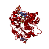
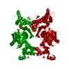
 Vibrio cholerae O1 biovar eltor (bacteria)
 X-RAY DIFFRACTION /
  Authors
 Citation
 Structure visualization
 Downloads & links
 Other downloads
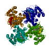
 PDBj
PDBj


 Assembly
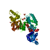


 Mass: 65.409 Da / Num. of mol.: 1 / Source method: obtained synthetically / Formula: Zn
 Mass: 96.063 Da / Num. of mol.: 6 / Source method: obtained synthetically / Formula: SO4
 Mass: 78.133 Da / Num. of mol.: 1 / Source method: obtained synthetically / Formula: C2H6OS
 Mass: 92.094 Da / Num. of mol.: 1 / Source method: obtained synthetically / Formula: C3H8O3
 Sample preparation
 / Beamline: 32-ID / Wavelength: 1 Å
 Processing