Movie
Movie Controller


+ Open data

- Basic information
| Entry | Database: EMDB / ID: EMD-24302 | |||||||||
|---|---|---|---|---|---|---|---|---|---|---|
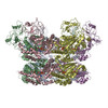
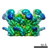
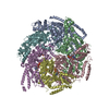
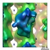
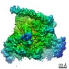
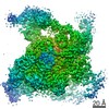
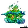
| Title | p47-bound p97-R155H mutant with ATPgammaS | |||||||||
 Map data Map data | p47-bound p97-R155H mutant with ATPgammaS | |||||||||
 Sample Sample |
| |||||||||
 Keywords Keywords | AAA+ ATPase / MOTOR PROTEIN / HYDROLASE / HYDROLASE-Lipid Binding Protein complex | |||||||||
| Function / homology |  Function and homology information Function and homology informationRHOH GTPase cycle / negative regulation of protein localization to centrosome / positive regulation of mitotic centrosome separation / nuclear membrane reassembly / spindle pole centrosome / flavin adenine dinucleotide catabolic process / VCP-NSFL1C complex / endoplasmic reticulum stress-induced pre-emptive quality control / endosome to lysosome transport via multivesicular body sorting pathway / Golgi stack ...RHOH GTPase cycle / negative regulation of protein localization to centrosome / positive regulation of mitotic centrosome separation / nuclear membrane reassembly / spindle pole centrosome / flavin adenine dinucleotide catabolic process / VCP-NSFL1C complex / endoplasmic reticulum stress-induced pre-emptive quality control / endosome to lysosome transport via multivesicular body sorting pathway / Golgi stack / BAT3 complex binding / cellular response to arsenite ion / cytoplasmic ubiquitin ligase complex / protein-DNA covalent cross-linking repair / Derlin-1 retrotranslocation complex / positive regulation of protein K63-linked deubiquitination / deubiquitinase activator activity / positive regulation of oxidative phosphorylation / cytoplasm protein quality control / aggresome assembly / ubiquitin-modified protein reader activity / regulation of protein localization to chromatin / cellular response to misfolded protein / mitotic spindle disassembly / VCP-NPL4-UFD1 AAA ATPase complex / positive regulation of mitochondrial membrane potential / vesicle-fusing ATPase / ATPase complex / K48-linked polyubiquitin modification-dependent protein binding / regulation of aerobic respiration / NAD+ metabolic process / retrograde protein transport, ER to cytosol / stress granule disassembly / Golgi organization / ubiquitin-specific protease binding / establishment of mitotic spindle orientation / regulation of synapse organization / ciliary transition zone / positive regulation of ATP biosynthetic process / intracellular membrane-bounded organelle / ubiquitin-like protein ligase binding / RHOH GTPase cycle / MHC class I protein binding / autophagosome maturation / negative regulation of hippo signaling / HSF1 activation / endoplasmic reticulum to Golgi vesicle-mediated transport / polyubiquitin modification-dependent protein binding / autophagosome assembly / ATP metabolic process / interstrand cross-link repair / Attachment and Entry / endoplasmic reticulum unfolded protein response / Protein methylation / ERAD pathway / ciliary tip / translesion synthesis / negative regulation of protein localization to chromatin / lipid droplet / viral genome replication / proteasome complex / Josephin domain DUBs / macroautophagy / proteasomal protein catabolic process / negative regulation of smoothened signaling pathway / ubiquitin binding / establishment of protein localization / N-glycan trimming in the ER and Calnexin/Calreticulin cycle / positive regulation of protein-containing complex assembly / ADP binding / Hh mutants are degraded by ERAD / Translesion Synthesis by POLH / positive regulation of non-canonical NF-kappaB signal transduction / Hedgehog ligand biogenesis / Defective CFTR causes cystic fibrosis / autophagy / ABC-family protein mediated transport / cytoplasmic stress granule / Aggrephagy / positive regulation of protein catabolic process / positive regulation of canonical Wnt signaling pathway / azurophil granule lumen / Ovarian tumor domain proteases / KEAP1-NFE2L2 pathway / double-strand break repair / positive regulation of proteasomal ubiquitin-dependent protein catabolic process / cellular response to heat / chromosome / E3 ubiquitin ligases ubiquitinate target proteins / site of double-strand break / ATPase binding / Neddylation / secretory granule lumen / protein phosphatase binding / regulation of apoptotic process / ficolin-1-rich granule lumen / ubiquitin-dependent protein catabolic process / membrane fusion / Attachment and Entry / proteasome-mediated ubiquitin-dependent protein catabolic process Similarity search - Function | |||||||||
| Biological species |  Homo sapiens (human) / Homo sapiens (human) /  | |||||||||
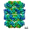
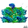
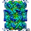
| Method | single particle reconstruction / cryo EM / Resolution: 4.23 Å | |||||||||
 Authors Authors | Nandi P / Li S | |||||||||
| Funding support |  United States, 1 items United States, 1 items
| |||||||||
 Citation Citation | Journal: Int J Mol Sci / Year: 2021 Title: Structural and Functional Analysis of Disease-Linked p97 ATPase Mutant Complexes. Authors: Purbasha Nandi / Shan Li / Rod Carlo A Columbres / Feng Wang / Dewight R Williams / Yu-Ping Poh / Tsui-Fen Chou / Po-Lin Chiu / Abstract: IBMPFD/ALS is a genetic disorder caused by a single amino acid mutation on the p97 ATPase, promoting ATPase activity and cofactor dysregulation. The disease mechanism underlying p97 ATPase ...IBMPFD/ALS is a genetic disorder caused by a single amino acid mutation on the p97 ATPase, promoting ATPase activity and cofactor dysregulation. The disease mechanism underlying p97 ATPase malfunction remains unclear. To understand how the mutation alters the ATPase regulation, we assembled a full-length p97 with its p47 cofactor and first visualized their structures using single-particle cryo-EM. More than one-third of the population was the dodecameric form. Nucleotide presence dissociates the dodecamer into two hexamers for its highly elevated function. The N-domains of the p97 mutant all show up configurations in ADP- or ATPS-bound states. Our functional and structural analyses showed that the p47 binding is likely to impact the p97 ATPase activities via changing the conformations of arginine fingers. These functional and structural analyses underline the ATPase dysregulation with the miscommunication between the functional modules of the p97. | |||||||||
| History |
|
- Structure visualization
| Movie |
Movie viewer |
|---|---|
| Structure viewer | EM map: SurfViewMolmilJmol/JSmol |
| Supplemental images |

- Downloads & links
-EMDB archive
| Map data | emd_24302.map.gz | 49.8 MB | EMDB map data format | |
|---|---|---|---|---|
| Header (meta data) | emd-24302-v30.xmlemd-24302.xml | 15.9 KB 15.9 KB | Display Display | EMDB header |
| Images | 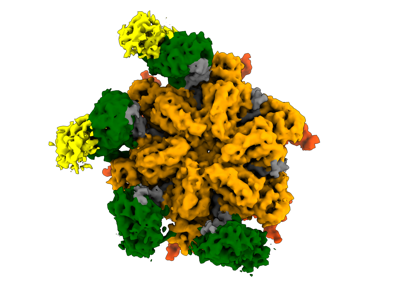 emd_24302.png emd_24302.png | 74.7 KB | ||
| Filedesc metadata | emd-24302.cif.gz | 6.7 KB | ||
| Archive directory |  http://ftp.pdbj.org/pub/emdb/structures/EMD-24302ftp://ftp.pdbj.org/pub/emdb/structures/EMD-24302 http://ftp.pdbj.org/pub/emdb/structures/EMD-24302ftp://ftp.pdbj.org/pub/emdb/structures/EMD-24302 | HTTPS FTP |
-Related structure data
| Related structure data |  7r7sMC  7l5wC  7l5xC  7r7tC 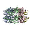 7r7uC M: atomic model generated by this map C: citing same article ( |
|---|---|
| Similar structure data | |
| EM raw data | EMPIAR-10920 (Title: Cryo-EM dataset of mutant p97R155H-p47 in presence of ATPγS collected using Thermo Fisher/FEI Titan Krios TEM Gatan K2 Summit DED camera Data size: 280.3 Data #1: Motion corrected dose-weighted micrographs for the cryo-EM dataset of disease mutant p97R155H-p47 in presence of ADP. [micrographs - single frame]) |


-Links
| EMDB pages | EMDB (EBI/PDBe) / EMDataResource |
|---|---|
| Related items in Molecule of the Month |


-Map
| File | Download / File: emd_24302.map.gz / Format: CCP4 / Size: 52.7 MB / Type: IMAGE STORED AS FLOATING POINT NUMBER (4 BYTES) | ||||||||||||||||||||||||||||||||||||||||||||||||||||||||||||||||||||
|---|---|---|---|---|---|---|---|---|---|---|---|---|---|---|---|---|---|---|---|---|---|---|---|---|---|---|---|---|---|---|---|---|---|---|---|---|---|---|---|---|---|---|---|---|---|---|---|---|---|---|---|---|---|---|---|---|---|---|---|---|---|---|---|---|---|---|---|---|---|
| Annotation | p47-bound p97-R155H mutant with ATPgammaS | ||||||||||||||||||||||||||||||||||||||||||||||||||||||||||||||||||||
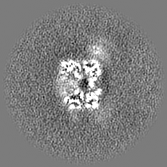
| Projections & slices | Image control
Images are generated by Spider. | ||||||||||||||||||||||||||||||||||||||||||||||||||||||||||||||||||||
| Voxel size | X=Y=Z: 1.413 Å | ||||||||||||||||||||||||||||||||||||||||||||||||||||||||||||||||||||
| Density |
| ||||||||||||||||||||||||||||||||||||||||||||||||||||||||||||||||||||
| Symmetry | Space group: 1 | ||||||||||||||||||||||||||||||||||||||||||||||||||||||||||||||||||||
| Details | EMDB XML:
CCP4 map header:
| ||||||||||||||||||||||||||||||||||||||||||||||||||||||||||||||||||||
 Z (Sec.)
 Y (Row.)
 X (Col.)


-Supplemental data
- Sample components
-Entire : p47-bound p97-R155H mutant with ATPgammaS
| Entire | Name: p47-bound p97-R155H mutant with ATPgammaS |
|---|---|
| Components |
|
-Supramolecule #1: p47-bound p97-R155H mutant with ATPgammaS
| Supramolecule | Name: p47-bound p97-R155H mutant with ATPgammaS / type: complex / ID: 1 / Parent: 0 / Macromolecule list: #1-#2 / Details: p47-bound p97-R155H mutant with ATPgammaS |
|---|---|
| Source (natural) | Organism: Homo sapiens (human) |
| Molecular weight | Theoretical: 723 KDa |
-Macromolecule #1: Transitional endoplasmic reticulum ATPase
| Macromolecule | Name: Transitional endoplasmic reticulum ATPase / type: protein_or_peptide / ID: 1 / Number of copies: 6 / Enantiomer: LEVO / EC number: vesicle-fusing ATPase |
|---|---|
| Source (natural) | Organism: Homo sapiens (human) |
| Molecular weight | Theoretical: 89.417773 KDa |
| Recombinant expression | Organism:  |
| Sequence | String: MASGADSKGD DLSTAILKQK NRPNRLIVDE AINEDNSVVS LSQPKMDELQ LFRGDTVLLK GKKRREAVCI VLSDDTCSDE KIRMNRVVR NNLRVRLGDV ISIQPCPDVK YGKRIHVLPI DDTVEGITGN LFEVYLKPYF LEAYRPIRKG DIFLVHGGMR A VEFKVVET ...String: MASGADSKGD DLSTAILKQK NRPNRLIVDE AINEDNSVVS LSQPKMDELQ LFRGDTVLLK GKKRREAVCI VLSDDTCSDE KIRMNRVVR NNLRVRLGDV ISIQPCPDVK YGKRIHVLPI DDTVEGITGN LFEVYLKPYF LEAYRPIRKG DIFLVHGGMR A VEFKVVET DPSPYCIVAP DTVIHCEGEP IKREDEEESL NEVGYDDIGG CRKQLAQIKE MVELPLRHPA LFKAIGVKPP RG ILLYGPP GTGKTLIARA VANETGAFFF LINGPEIMSK LAGESESNLR KAFEEAEKNA PAIIFIDELD AIAPKREKTH GEV ERRIVS QLLTLMDGLK QRAHVIVMAA TNRPNSIDPA LRRFGRFDRE VDIGIPDATG RLEILQIHTK NMKLADDVDL EQVA NETHG HVGADLAALC SEAALQAIRK KMDLIDLEDE TIDAEVMNSL AVTMDDFRWA LSQSNPSALR ETVVEVPQVT WEDIG GLED VKRELQELVQ YPVEHPDKFL KFGMTPSKGV LFYGPPGCGK TLLAKAIANE CQANFISIKG PELLTMWFGE SEANVR EIF DKARQAAPCV LFFDELDSIA KARGGNIGDG GGAADRVINQ ILTEMDGMST KKNVFIIGAT NRPDIIDPAI LRPGRLD QL IYIPLPDEKS RVAILKANLR KSPVAKDVDL EFLAKMTNGF SGADLTEICQ RACKLAIRES IESEIRRERE RQTNPSAM E VEEDDPVPEI RRDHFEEAMR FARRSVSDND IRKYEMFAQT LQQSRGFGSF RFPSGNQGGA GPSQGSGGGT GGSVYTEDN DDDLYG UniProtKB: Transitional endoplasmic reticulum ATPase |
-Macromolecule #2: NSFL1 cofactor p47
| Macromolecule | Name: NSFL1 cofactor p47 / type: protein_or_peptide / ID: 2 / Number of copies: 2 / Enantiomer: LEVO |
|---|---|
| Source (natural) | Organism: |
| Molecular weight | Theoretical: 40.731855 KDa |
| Recombinant expression | Organism: |
| Sequence | String: MAEERQDALR EFVAVTGAEE DRARFFLESA GWDLQIALAS FYEDGGDEDI VTISQATPSS VSRGTAPSDN RVTSFRDLIH DQDEEEEEE EGQRFYAGGS ERSGQQIVGP PRKKSPNELV DDLFKGAKEH GAVAVERVTK SPGETSKPRP FAGGGYRLGA A PEEESAYV ...String: MAEERQDALR EFVAVTGAEE DRARFFLESA GWDLQIALAS FYEDGGDEDI VTISQATPSS VSRGTAPSDN RVTSFRDLIH DQDEEEEEE EGQRFYAGGS ERSGQQIVGP PRKKSPNELV DDLFKGAKEH GAVAVERVTK SPGETSKPRP FAGGGYRLGA A PEEESAYV AGERRRHSGQ DVHVVLKLWK TGFSLDNGDL RSYQDPSNAQ FLESIRRGEV PAELRRLAHG GQVNLDMEDH RD EDFVKPK GAFKAFTGEG QKLGSTAPQV LNTSSPAQQA ENEAKASSSI LINEAEPTTN IQIRLADGGR LVQKFNHSHR ISD IRLFIV DARPAMAATS FVLMTTFPNK ELADENQTLK EANLLNAVIV QRLT UniProtKB: NSFL1 cofactor p47 |
-Macromolecule #3: PHOSPHOTHIOPHOSPHORIC ACID-ADENYLATE ESTER
| Macromolecule | Name: PHOSPHOTHIOPHOSPHORIC ACID-ADENYLATE ESTER / type: ligand / ID: 3 / Number of copies: 12 / Formula: AGS |
|---|---|
| Molecular weight | Theoretical: 523.247 Da |
| Chemical component information |  ChemComp-AGS: |
-Experimental details
-Structure determination
| Method | cryo EM |
|---|---|
 Processing Processing | single particle reconstruction |
| Aggregation state | particle |
-Sample preparation
| Concentration | 0.2 mg/mL | ||||||||||||
|---|---|---|---|---|---|---|---|---|---|---|---|---|---|
| Buffer | pH: 7.4 Component:
| ||||||||||||
| Grid | Model: C-flat-2/1 / Support film - Material: CARBON / Support film - topology: HOLEY ARRAY | ||||||||||||
| Vitrification | Cryogen name: ETHANE / Chamber humidity: 100 % / Chamber temperature: 292 K / Instrument: FEI VITROBOT MARK IV / Details: The grid was blotted for 6 seconds.. | ||||||||||||
| Details | The protein sample was monodisperse. |
- Electron microscopy
| Microscope | FEI TITAN KRIOS |
|---|---|
| Alignment procedure | Coma free - Residual tilt: 0.001 mrad |
| Image recording | Film or detector model: GATAN K2 SUMMIT (4k x 4k) / Detector mode: SUPER-RESOLUTION / Number real images: 2796 / Average electron dose: 44.4 e/Å2 |
| Electron beam | Acceleration voltage: 300 kV / Electron source:  FIELD EMISSION GUN FIELD EMISSION GUN |
| Electron optics | C2 aperture diameter: 50.0 µm / Illumination mode: FLOOD BEAM / Imaging mode: BRIGHT FIELD / Cs: 2.7 mm / Nominal defocus max: -2.5 µm / Nominal defocus min: -0.8 µm / Nominal magnification: 48077 |
| Sample stage | Specimen holder model: FEI TITAN KRIOS AUTOGRID HOLDER / Cooling holder cryogen: NITROGEN |
| Experimental equipment |  Model: Titan Krios / Image courtesy: FEI Company |