 Movie
Movie Controller


+ Open data

- Basic information
| Entry | Database: PDB / ID: 5ftn | ||||||
|---|---|---|---|---|---|---|---|
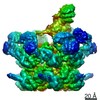
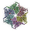
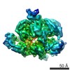
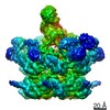
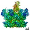
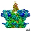
| Title | Cryo-EM structure of human p97 bound to ATPgS (Conformation III) | ||||||
 Components Components | TRANSITIONAL ENDOPLASMIC RETICULUM ATPASE | ||||||
 Keywords Keywords | HYDROLASE / SINGLE-PARTICLE / P97 / AAA ATPASE | ||||||
| Function / homology |  Function and homology information Function and homology informationflavin adenine dinucleotide catabolic process / VCP-NSFL1C complex / endoplasmic reticulum stress-induced pre-emptive quality control / endosome to lysosome transport via multivesicular body sorting pathway / BAT3 complex binding / cellular response to arsenite ion / cytoplasmic ubiquitin ligase complex / protein-DNA covalent cross-linking repair / Derlin-1 retrotranslocation complex / positive regulation of protein K63-linked deubiquitination ...flavin adenine dinucleotide catabolic process / VCP-NSFL1C complex / endoplasmic reticulum stress-induced pre-emptive quality control / endosome to lysosome transport via multivesicular body sorting pathway / BAT3 complex binding / cellular response to arsenite ion / cytoplasmic ubiquitin ligase complex / protein-DNA covalent cross-linking repair / Derlin-1 retrotranslocation complex / positive regulation of protein K63-linked deubiquitination / ATPase complex / deubiquitinase activator activity / positive regulation of oxidative phosphorylation / cytoplasm protein quality control / aggresome assembly / ubiquitin-modified protein reader activity / regulation of protein localization to chromatin / cellular response to misfolded protein / mitotic spindle disassembly / VCP-NPL4-UFD1 AAA ATPase complex / positive regulation of mitochondrial membrane potential / vesicle-fusing ATPase / K48-linked polyubiquitin modification-dependent protein binding / ciliary transition zone / regulation of aerobic respiration / NAD+ metabolic process / retrograde protein transport, ER to cytosol / stress granule disassembly / ubiquitin-specific protease binding / regulation of synapse organization / positive regulation of ATP biosynthetic process / intracellular membrane-bounded organelle / ubiquitin-like protein ligase binding / RHOH GTPase cycle / MHC class I protein binding / autophagosome maturation / negative regulation of hippo signaling / endoplasmic reticulum to Golgi vesicle-mediated transport / HSF1 activation / polyubiquitin modification-dependent protein binding / interstrand cross-link repair / ATP metabolic process / Attachment and Entry / endoplasmic reticulum unfolded protein response / Protein methylation / ERAD pathway / ciliary tip / translesion synthesis / negative regulation of protein localization to chromatin / lipid droplet / viral genome replication / proteasome complex / macroautophagy / negative regulation of smoothened signaling pathway / Josephin domain DUBs / establishment of protein localization / proteasomal protein catabolic process / N-glycan trimming in the ER and Calnexin/Calreticulin cycle / positive regulation of protein-containing complex assembly / ADP binding / Hh mutants are degraded by ERAD / positive regulation of non-canonical NF-kappaB signal transduction / Translesion Synthesis by POLH / Hedgehog ligand biogenesis / Defective CFTR causes cystic fibrosis / ABC-family protein mediated transport / autophagy / cytoplasmic stress granule / positive regulation of protein catabolic process / positive regulation of canonical Wnt signaling pathway / Aggrephagy / azurophil granule lumen / Ovarian tumor domain proteases / KEAP1-NFE2L2 pathway / double-strand break repair / positive regulation of proteasomal ubiquitin-dependent protein catabolic process / cellular response to heat / E3 ubiquitin ligases ubiquitinate target proteins / site of double-strand break / Neddylation / secretory granule lumen / protein phosphatase binding / regulation of apoptotic process / ficolin-1-rich granule lumen / ciliary basal body / ubiquitin-dependent protein catabolic process / proteasome-mediated ubiquitin-dependent protein catabolic process / Attachment and Entry / protein ubiquitination / protein domain specific binding / DNA repair / ubiquitin protein ligase binding / Neutrophil degranulation / DNA damage response / lipid binding / endoplasmic reticulum membrane / perinuclear region of cytoplasm / glutamatergic synapse / endoplasmic reticulum / ATP hydrolysis activity Similarity search - Function | ||||||
| Biological species |  HOMO SAPIENS (human) HOMO SAPIENS (human) | ||||||
| Method | ELECTRON MICROSCOPY / single particle reconstruction / cryo EM / Resolution: 3.3 Å | ||||||
 Authors Authors | Banerjee, S. / Bartesaghi, A. / Merk, A. / Rao, P. / Bulfer, S.L. / Yan, Y. / Green, N. / Mroczkowski, B. / Neitz, R.J. / Wipf, P. ...Banerjee, S. / Bartesaghi, A. / Merk, A. / Rao, P. / Bulfer, S.L. / Yan, Y. / Green, N. / Mroczkowski, B. / Neitz, R.J. / Wipf, P. / Falconieri, V. / Deshaies, R.J. / Milne, J.L.S. / Huryn, D. / Arkin, M. / Subramaniam, S. | ||||||
 Citation Citation | Journal: Science / Year: 2016 Title: 2.3 Å resolution cryo-EM structure of human p97 and mechanism of allosteric inhibition. Authors: Soojay Banerjee / Alberto Bartesaghi / Alan Merk / Prashant Rao / Stacie L Bulfer / Yongzhao Yan / Neal Green / Barbara Mroczkowski / R Jeffrey Neitz / Peter Wipf / Veronica Falconieri / ...Authors: Soojay Banerjee / Alberto Bartesaghi / Alan Merk / Prashant Rao / Stacie L Bulfer / Yongzhao Yan / Neal Green / Barbara Mroczkowski / R Jeffrey Neitz / Peter Wipf / Veronica Falconieri / Raymond J Deshaies / Jacqueline L S Milne / Donna Huryn / Michelle Arkin / Sriram Subramaniam /  Abstract: p97 is a hexameric AAA+ adenosine triphosphatase (ATPase) that is an attractive target for cancer drug development. We report cryo-electron microscopy (cryo-EM) structures for adenosine diphosphate ...p97 is a hexameric AAA+ adenosine triphosphatase (ATPase) that is an attractive target for cancer drug development. We report cryo-electron microscopy (cryo-EM) structures for adenosine diphosphate (ADP)-bound, full-length, hexameric wild-type p97 in the presence and absence of an allosteric inhibitor at resolutions of 2.3 and 2.4 angstroms, respectively. We also report cryo-EM structures (at resolutions of ~3.3, 3.2, and 3.3 angstroms, respectively) for three distinct, coexisting functional states of p97 with occupancies of zero, one, or two molecules of adenosine 5'-O-(3-thiotriphosphate) (ATPγS) per protomer. A large corkscrew-like change in molecular architecture, coupled with upward displacement of the N-terminal domain, is observed only when ATPγS is bound to both the D1 and D2 domains of the protomer. These cryo-EM structures establish the sequence of nucleotide-driven structural changes in p97 at atomic resolution. They also enable elucidation of the binding mode of an allosteric small-molecule inhibitor to p97 and illustrate how inhibitor binding at the interface between the D1 and D2 domains prevents propagation of the conformational changes necessary for p97 function. | ||||||
| History |
|
- Structure visualization
| Movie |
Movie viewer |
|---|---|
| Structure viewer | Molecule: MolmilJmol/JSmol |
- Downloads & links
-Download
| PDBx/mmCIF format | 5ftn.cif.gz | 846.9 KB | Display | PDBx/mmCIF format |
|---|---|---|---|---|
| PDB format | pdb5ftn.ent.gz | 703.7 KB | Display | PDB format |
| PDBx/mmJSON format | 5ftn.json.gz | Tree view | PDBx/mmJSON format | |
| Others |  Other downloads Other downloads |
-Validation report
| Arichive directory | https://data.pdbj.org/pub/pdb/validation_reports/ft/5ftnftp://data.pdbj.org/pub/pdb/validation_reports/ft/5ftn | HTTPS FTP |
|---|
-Related structure data
| Related structure data | 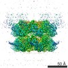 3299MC 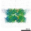 3295C 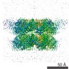 3296C 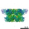 3297C 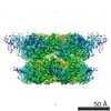 3298C 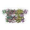 5ftjC 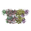 5ftkC 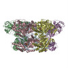 5ftlC 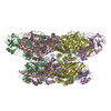 5ftmC C: citing same article ( M: map data used to model this data |
|---|---|
| Similar structure data |
-Links
 PDBj
PDBj


- Assembly
| Deposited unit | 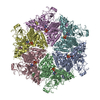
|
|---|---|
| 1 |
|
-Components
| #1: Protein | Mass: 89436.820 Da / Num. of mol.: 6 Source method: isolated from a genetically manipulated source Source: (gene. exp.) HOMO SAPIENS (human) / Production host:  #2: Chemical | ChemComp-AGS /   Mass: 523.247 Da / Num. of mol.: 12 / Source method: obtained synthetically / Formula: C10H16N5O12P3S / Comment: ATP-gamma-S, energy-carrying molecule analogue*YM Mass: 523.247 Da / Num. of mol.: 12 / Source method: obtained synthetically / Formula: C10H16N5O12P3S / Comment: ATP-gamma-S, energy-carrying molecule analogue*YM#3: Chemical | ChemComp-MG /   Mass: 24.305 Da / Num. of mol.: 12 / Source method: obtained synthetically / Formula: Mg Mass: 24.305 Da / Num. of mol.: 12 / Source method: obtained synthetically / Formula: Mg |
|---|
-Experimental details
-Experiment
| Experiment | Method: ELECTRON MICROSCOPY |
|---|---|
| EM experiment | Aggregation state: PARTICLE / 3D reconstruction method: single particle reconstruction |
- Sample preparation
| Component | Name: FULL-LENGTH HUMAN P97 BOUND TO ATPGS / Type: COMPLEX |
|---|---|
| Buffer solution | Name: 25 MM TRIS, 150 MM NACL, 1 MM MGCL2, 1.0 MM TCEP / pH: 8 / Details: 25 MM TRIS, 150 MM NACL, 1 MM MGCL2, 1.0 MM TCEP |
| Specimen | Conc.: 0.9 mg/ml / Embedding applied: NO / Shadowing applied: NO / Staining applied: NO / Vitrification applied: YES |
| Specimen support | Details: HOLEY CARBON |
| Vitrification | Instrument: FEI VITROBOT MARK IV / Cryogen name: ETHANE Details: VITRIFICATION 1 -- CRYOGEN- ETHANE, HUMIDITY- 100, TEMPERATURE- 90.15, INSTRUMENT- FEI VITROBOT MARK IV, METHOD- BLOT FOR 2.5 SECONDS BEFORE PLUNGING. |
- Electron microscopy imaging
| Experimental equipment |  Model: Titan Krios / Image courtesy: FEI Company |
|---|---|
| Microscopy | Model: FEI TITAN KRIOS / Date: Jan 17, 2015 / Details: PARALLEL BEAM ILLUMINATION |
| Electron gun | Electron source:  FIELD EMISSION GUN / Accelerating voltage: 300 kV / Illumination mode: FLOOD BEAM FIELD EMISSION GUN / Accelerating voltage: 300 kV / Illumination mode: FLOOD BEAM |
| Electron lens | Mode: BRIGHT FIELD / Nominal magnification: 105000 X / Calibrated magnification: 36980 X / Nominal defocus max: 2700 nm / Nominal defocus min: 950 nm / Cs: 2.7 mm |
| Specimen holder | Temperature: 79.7 K |
| Image recording | Electron dose: 40 e/Å2 / Film or detector model: GATAN K2 SUMMIT (4k x 4k) |
| Image scans | Num. digital images: 628 |
- Processing
| EM software | Name: FREALIGN / Category: 3D reconstruction | ||||||||||||
|---|---|---|---|---|---|---|---|---|---|---|---|---|---|
| CTF correction | Details: EACH PARTICLE | ||||||||||||
| Symmetry | Point symmetry: C6 (6 fold cyclic) | ||||||||||||
| 3D reconstruction | Method: SCORE MINIMIZATION / Resolution: 3.3 Å / Num. of particles: 23306 Details: N-TERMINAL RESIDUES 21-200 DISORDERED. LINKER CONNECTING RESIDUES 707 TO 728 IS NOT IN THE MODEL. SUBMISSION BASED ON EXPERIMENTAL DATA FROM EMDB EMD-3299. (DEPOSITION ID: 14179). Symmetry type: POINT | ||||||||||||
| Refinement | Highest resolution: 3.3 Å | ||||||||||||
| Refinement step | Cycle: LAST / Highest resolution: 3.3 Å
|
