 Movie
Movie Controller

[English] 日本語
 Yorodumi
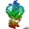
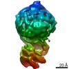
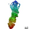
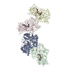
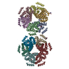
- EMDB-23980: Rhodopsin kinase (GRK1)-S5E/S488E/T489E in complex with rhodopsin... -
+ Open data

- Basic information
| Entry | Database: EMDB / ID: EMD-23980 | |||||||||||||||
|---|---|---|---|---|---|---|---|---|---|---|---|---|---|---|---|---|
| Title | Rhodopsin kinase (GRK1)-S5E/S488E/T489E in complex with rhodopsin and Fab6 | |||||||||||||||
 Map data Map data | Rhodopsin kinase (GRK1)-S5E/S488E/T489E in complex with rhodopsin and Fab6 | |||||||||||||||
 Sample Sample |
| |||||||||||||||
 Keywords Keywords | GPCR / signal desensitization / Kinase / MEMBRANE PROTEIN / MEMBRANE PROTEIN-Immune System complex / MEMBRANE / Signaling protein-Immune System complex | |||||||||||||||
| Function / homology |  Function and homology information Function and homology informationrhodopsin kinase / rhodopsin kinase activity / Opsins / VxPx cargo-targeting to cilium / rod bipolar cell differentiation / sperm head plasma membrane / regulation of opsin-mediated signaling pathway / opsin binding / The canonical retinoid cycle in rods (twilight vision) / absorption of visible light ...rhodopsin kinase / rhodopsin kinase activity / Opsins / VxPx cargo-targeting to cilium / rod bipolar cell differentiation / sperm head plasma membrane / regulation of opsin-mediated signaling pathway / opsin binding / The canonical retinoid cycle in rods (twilight vision) / absorption of visible light / G protein-coupled opsin signaling pathway / 11-cis retinal binding / podosome assembly / G protein-coupled photoreceptor activity / photoreceptor inner segment membrane / rod photoreceptor outer segment / cellular response to light stimulus / G protein-coupled receptor complex / Inactivation, recovery and regulation of the phototransduction cascade / thermotaxis / Activation of the phototransduction cascade / detection of temperature stimulus involved in thermoception / outer membrane / response to light intensity / photoreceptor cell maintenance / arrestin family protein binding / photoreceptor outer segment membrane / G alpha (i) signalling events / phototransduction, visible light / phototransduction / response to light stimulus / regulation of signal transduction / photoreceptor outer segment / G-protein alpha-subunit binding / visual perception / guanyl-nucleotide exchange factor activity / microtubule cytoskeleton organization / cell-cell junction / photoreceptor disc membrane / protein autophosphorylation / sperm midpiece / gene expression / G protein-coupled receptor signaling pathway / Golgi membrane / signal transduction / zinc ion binding / ATP binding / membrane / identical protein binding / plasma membrane / cytoplasm Similarity search - Function | |||||||||||||||
| Biological species |  Homo sapiens (human) / Homo sapiens (human) /  | |||||||||||||||
| Method | single particle reconstruction / cryo EM / Resolution: 4.0 Å | |||||||||||||||
 Authors Authors | Chen Q / Tesmer JJG | |||||||||||||||
| Funding support |  United States, 4 items United States, 4 items
| |||||||||||||||
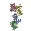
 Citation Citation | Journal: Nature / Year: 2021 Title: Structures of rhodopsin in complex with G-protein-coupled receptor kinase 1. Authors: Qiuyan Chen / Manolo Plasencia / Zhuang Li / Somnath Mukherjee / Dhabaleswar Patra / Chun-Liang Chen / Thomas Klose / Xin-Qiu Yao / Anthony A Kossiakoff / Leifu Chang / Philip C Andrews / John J G Tesmer / Abstract: G-protein-coupled receptor (GPCR) kinases (GRKs) selectively phosphorylate activated GPCRs, thereby priming them for desensitization. Although it is unclear how GRKs recognize these receptors, a ...G-protein-coupled receptor (GPCR) kinases (GRKs) selectively phosphorylate activated GPCRs, thereby priming them for desensitization. Although it is unclear how GRKs recognize these receptors, a conserved region at the GRK N terminus is essential for this process. Here we report a series of cryo-electron microscopy single-particle reconstructions of light-activated rhodopsin (Rho*) bound to rhodopsin kinase (GRK1), wherein the N terminus of GRK1 forms a helix that docks into the open cytoplasmic cleft of Rho*. The helix also packs against the GRK1 kinase domain and stabilizes it in an active configuration. The complex is further stabilized by electrostatic interactions between basic residues that are conserved in most GPCRs and acidic residues that are conserved in GRKs. We did not observe any density for the regulator of G-protein signalling homology domain of GRK1 or the C terminus of rhodopsin. Crosslinking with mass spectrometry analysis confirmed these results and revealed dynamic behaviour in receptor-bound GRK1 that would allow the phosphorylation of multiple sites in the receptor tail. We have identified GRK1 residues whose mutation augments kinase activity and crosslinking with Rho*, as well as residues that are involved in activation by acidic phospholipids. From these data, we present a general model for how a small family of protein kinases can recognize and be activated by hundreds of different GPCRs. | |||||||||||||||
| History |
|
- Structure visualization
| Movie |
Movie viewer |
|---|---|
| Structure viewer | EM map: SurfViewMolmilJmol/JSmol |
| Supplemental images |

- Downloads & links
-EMDB archive
| Map data | emd_23980.map.gz | 230.3 MB | EMDB map data format | |
|---|---|---|---|---|
| Header (meta data) | emd-23980-v30.xmlemd-23980.xml | 15.9 KB 15.9 KB | Display Display | EMDB header |
| FSC (resolution estimation) | emd_23980_fsc.xml | 13.9 KB | Display | FSC data file |
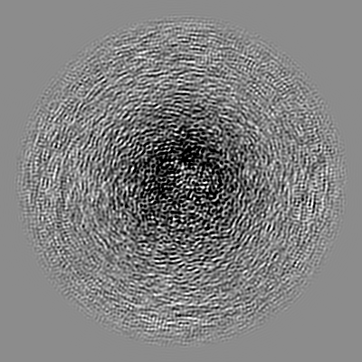
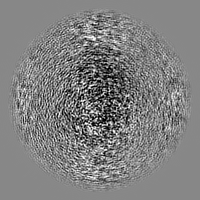
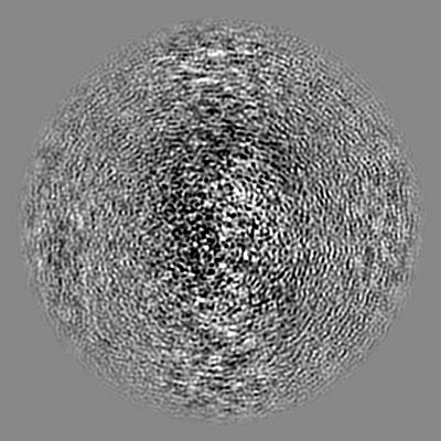
| Images | 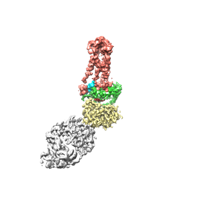 emd_23980.png emd_23980.png | 49.4 KB | ||
| Filedesc metadata | emd-23980.cif.gz | 6.4 KB | ||
| Archive directory |  http://ftp.pdbj.org/pub/emdb/structures/EMD-23980ftp://ftp.pdbj.org/pub/emdb/structures/EMD-23980 http://ftp.pdbj.org/pub/emdb/structures/EMD-23980ftp://ftp.pdbj.org/pub/emdb/structures/EMD-23980 | HTTPS FTP |
-Related structure data
| Related structure data | 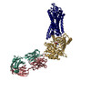 7mtbMC  7mt8C 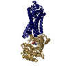 7mt9C 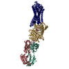 7mtaC M: atomic model generated by this map C: citing same article ( |
|---|---|
| Similar structure data |


-Links
| EMDB pages | EMDB (EBI/PDBe) / EMDataResource |
|---|---|
| Related items in Molecule of the Month |


-Map
| File | Download / File: emd_23980.map.gz / Format: CCP4 / Size: 244.1 MB / Type: IMAGE STORED AS FLOATING POINT NUMBER (4 BYTES) | ||||||||||||||||||||||||||||||||||||||||||||||||||||||||||||||||||||
|---|---|---|---|---|---|---|---|---|---|---|---|---|---|---|---|---|---|---|---|---|---|---|---|---|---|---|---|---|---|---|---|---|---|---|---|---|---|---|---|---|---|---|---|---|---|---|---|---|---|---|---|---|---|---|---|---|---|---|---|---|---|---|---|---|---|---|---|---|---|
| Annotation | Rhodopsin kinase (GRK1)-S5E/S488E/T489E in complex with rhodopsin and Fab6 | ||||||||||||||||||||||||||||||||||||||||||||||||||||||||||||||||||||
| Projections & slices | Image control
Images are generated by Spider. | ||||||||||||||||||||||||||||||||||||||||||||||||||||||||||||||||||||
| Voxel size | X=Y=Z: 1.08 Å | ||||||||||||||||||||||||||||||||||||||||||||||||||||||||||||||||||||
| Density |
| ||||||||||||||||||||||||||||||||||||||||||||||||||||||||||||||||||||
| Symmetry | Space group: 1 | ||||||||||||||||||||||||||||||||||||||||||||||||||||||||||||||||||||
| Details | EMDB XML:
CCP4 map header:
| ||||||||||||||||||||||||||||||||||||||||||||||||||||||||||||||||||||
 Z (Sec.)
 Y (Row.)
 X (Col.)


-Supplemental data
- Sample components
-Entire : Rhodopsin kinase (GRK1)-S5E/S488E/T489E in complex with rhodopsin...
| Entire | Name: Rhodopsin kinase (GRK1)-S5E/S488E/T489E in complex with rhodopsin stabilized by Fab6 |
|---|---|
| Components |
|
-Supramolecule #1: Rhodopsin kinase (GRK1)-S5E/S488E/T489E in complex with rhodopsin...
| Supramolecule | Name: Rhodopsin kinase (GRK1)-S5E/S488E/T489E in complex with rhodopsin stabilized by Fab6 type: complex / ID: 1 / Parent: 0 / Macromolecule list: #1-#4 |
|---|
-Supramolecule #2: Fab6 Heavy and Light Chains
| Supramolecule | Name: Fab6 Heavy and Light Chains / type: complex / ID: 2 / Parent: 1 / Macromolecule list: #2-#3 |
|---|---|
| Source (natural) | Organism: Homo sapiens (human) |
-Supramolecule #3: Rhodopsin kinase, rhodopsin
| Supramolecule | Name: Rhodopsin kinase, rhodopsin / type: complex / ID: 3 / Parent: 1 / Macromolecule list: #1, #4 |
|---|---|
| Source (natural) | Organism: |
-Macromolecule #1: Rhodopsin kinase GRK1
| Macromolecule | Name: Rhodopsin kinase GRK1 / type: protein_or_peptide / ID: 1 / Number of copies: 1 / Enantiomer: LEVO / EC number: rhodopsin kinase |
|---|---|
| Source (natural) | Organism: |
| Molecular weight | Theoretical: 61.542012 KDa |
| Recombinant expression | Organism:   Spodoptera frugiperda (fall armyworm) Spodoptera frugiperda (fall armyworm) |
| Sequence | String: MDFGELETVV ANSAFIAARG SFDASSGPAS RDRKYLARLK LPPLSKCEAL RESLDLGFEG MCLEQPIGKR LFQQFLRTHE QHGPALQLW KDIEDYDTAD DALRPQKAQA LRAAYLEPQA QLFCSFLDAE TVARARAGAG DGLFQPLLRA VLAHLGQAPF Q EFLDSLYF ...String: MDFGELETVV ANSAFIAARG SFDASSGPAS RDRKYLARLK LPPLSKCEAL RESLDLGFEG MCLEQPIGKR LFQQFLRTHE QHGPALQLW KDIEDYDTAD DALRPQKAQA LRAAYLEPQA QLFCSFLDAE TVARARAGAG DGLFQPLLRA VLAHLGQAPF Q EFLDSLYF LRFLQWKWLE AQPMGEDWFL DFRVLGRGGF GEVFACQMKA TGKLYACKKL NKKRLKKRKG YQGAMVEKKI LA KVHSRFI VSLAYAFETK TDLCLVMTIM NGGDIRYHIY NVDEDNPGFQ EPRAIFYTAQ IVSGLEHLHQ RNIIYRDLKP ENV LLDDDG NVRISDLGLA VELKAGQTKT KGYAGTPGFM APELLLGEEY DFSVDYFALG VTLYEMIAAR GPFRARGEKV ENKE LKQRV LEQAVTYPDK FSPASKDFCE ALLQKDPEKR LGFRDGSCDG LRTHPLFRDI SWRQLEAGML TPPFVPDSRT VYAKN IQDV GAFEEVKGVA FEKADTEFFQ EFASGTCPIP WQEEMIETGV FGDLNVWRPD GVDHHHHHH UniProtKB: Rhodopsin kinase GRK1 |
-Macromolecule #2: Fab6 heavy chain
| Macromolecule | Name: Fab6 heavy chain / type: protein_or_peptide / ID: 2 / Number of copies: 1 / Enantiomer: LEVO |
|---|---|
| Source (natural) | Organism: Homo sapiens (human) |
| Molecular weight | Theoretical: 25.170939 KDa |
| Recombinant expression | Organism:  |
| Sequence | String: EISEVQLVES GGGLVQPGGS LRLSCAASGF NFSSYSIHWV RQAPGKGLEW VAYISSYYGS TYYADSVKGR FTISADTSKN TAYLQMNSL RAEDTAVYYC ARHEYGWWND RWGLDYWGQG TLVTVSSAST KGPSVFPLAP SSKSTSGGTA ALGCLVKDYF P EPVTVSWN ...String: EISEVQLVES GGGLVQPGGS LRLSCAASGF NFSSYSIHWV RQAPGKGLEW VAYISSYYGS TYYADSVKGR FTISADTSKN TAYLQMNSL RAEDTAVYYC ARHEYGWWND RWGLDYWGQG TLVTVSSAST KGPSVFPLAP SSKSTSGGTA ALGCLVKDYF P EPVTVSWN SGALTSGVHT FPAVLQSSGL YSLSSVVTVP SSSLGTQTYI CNVNHKPSNT KVDKKVEPKS CDKTHT |
-Macromolecule #3: Fab6 light chain
| Macromolecule | Name: Fab6 light chain / type: protein_or_peptide / ID: 3 / Number of copies: 1 / Enantiomer: LEVO |
|---|---|
| Source (natural) | Organism: Homo sapiens (human) |
| Molecular weight | Theoretical: 23.509039 KDa |
| Recombinant expression | Organism: |
| Sequence | String: SDIQMTQSPS SLSASVGDRV TITCRASQSV SSAVAWYQQK PGKAPKLLIY SASSLYSGVP SRFSGSRSGT DFTLTISSLQ PEDFATYYC QQSSWYGPFT FGQGTKVEIK RTVAAPSVFI FPPSDSQLKS GTASVVCLLN NFYPREAKVQ WKVDNALQSG N SQESVTEQ ...String: SDIQMTQSPS SLSASVGDRV TITCRASQSV SSAVAWYQQK PGKAPKLLIY SASSLYSGVP SRFSGSRSGT DFTLTISSLQ PEDFATYYC QQSSWYGPFT FGQGTKVEIK RTVAAPSVFI FPPSDSQLKS GTASVVCLLN NFYPREAKVQ WKVDNALQSG N SQESVTEQ DSKDSTYSLS STLTLSKADY EKHKVYACEV THQGLSSPVT KSFNRGEC |
-Macromolecule #4: Rhodopsin
| Macromolecule | Name: Rhodopsin / type: protein_or_peptide / ID: 4 / Number of copies: 1 / Enantiomer: LEVO |
|---|---|
| Source (natural) | Organism: |
| Molecular weight | Theoretical: 39.031457 KDa |
| Sequence | String: MNGTEGPNFY VPFSNKTGVV RSPFEAPQYY LAEPWQFSML AAYMFLLIML GFPINFLTLY VTVQHKKLRT PLNYILLNLA VADLFMVFG GFTTTLYTSL HGYFVFGPTG CNLEGFFATL GGEIALWSLV VLAIERYVVV CKPMSNFRFG ENHAIMGVAF T WVMALACA ...String: MNGTEGPNFY VPFSNKTGVV RSPFEAPQYY LAEPWQFSML AAYMFLLIML GFPINFLTLY VTVQHKKLRT PLNYILLNLA VADLFMVFG GFTTTLYTSL HGYFVFGPTG CNLEGFFATL GGEIALWSLV VLAIERYVVV CKPMSNFRFG ENHAIMGVAF T WVMALACA APPLVGWSRY IPEGMQCSCG IDYYTPHEET NNESFVIYMF VVHFIIPLIV IFFCYGQLVF TVKEAAAQQQ ES ATTQKAE KEVTRMVIIM VIAFLICWLP YAGVAFYIFT HQGSDFGPIF MTIPAFFAKT SAVYNPVIYI MMNKQFRNCM VTT LCCGKN PLGDDEASTT VSKTETSQVA PA UniProtKB: Rhodopsin |
-Macromolecule #5: SANGIVAMYCIN
| Macromolecule | Name: SANGIVAMYCIN / type: ligand / ID: 5 / Number of copies: 1 / Formula: SGV |
|---|---|
| Molecular weight | Theoretical: 309.278 Da |
| Chemical component information |  ChemComp-SGV: |
-Macromolecule #6: RETINAL
| Macromolecule | Name: RETINAL / type: ligand / ID: 6 / Number of copies: 1 / Formula: RET |
|---|---|
| Molecular weight | Theoretical: 284.436 Da |
| Chemical component information |  ChemComp-RET: |
-Experimental details
-Structure determination
| Method | cryo EM |
|---|---|
 Processing Processing | single particle reconstruction |
| Aggregation state | particle |
-Sample preparation
| Buffer | pH: 8 |
|---|---|
| Vitrification | Cryogen name: ETHANE |
- Electron microscopy
| Microscope | FEI TITAN KRIOS |
|---|---|
| Image recording | Film or detector model: GATAN K3 BIOQUANTUM (6k x 4k) / Average electron dose: 54.0 e/Å2 |
| Electron beam | Acceleration voltage: 300 kV / Electron source:  FIELD EMISSION GUN FIELD EMISSION GUN |
| Electron optics | Illumination mode: FLOOD BEAM / Imaging mode: BRIGHT FIELD |
| Experimental equipment |  Model: Titan Krios / Image courtesy: FEI Company |

