Movie
Movie Controller


+ Open data

- Basic information
| Entry | Database: EMDB / ID: EMD-23813 | |||||||||
|---|---|---|---|---|---|---|---|---|---|---|
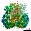
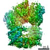
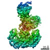
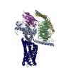
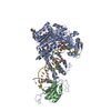
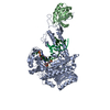
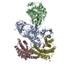
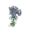
| Title | Autoinhibited B-Raf:(14-3-3)2:MEK complex with resolved RBD | |||||||||
 Map data Map data | ||||||||||
 Sample Sample |
| |||||||||
 Keywords Keywords | B-Raf / MEK / 14-3-3 / B-Raf complex / B-Raf monomer / Inactive B-Raf / Serine/threonine-protein kinase B-raf / RBD / signaling protein / SIGNALING PROTEIN-TRANSFERASE complex | |||||||||
| Function / homology |  Function and homology information Function and homology informationsynaptic target recognition / negative regulation of homotypic cell-cell adhesion / Golgi reassembly / negative regulation of hypoxia-induced intrinsic apoptotic signaling pathway / regulation of vascular associated smooth muscle contraction / CD4-positive, alpha-beta T cell differentiation / NOTCH4 Activation and Transmission of Signal to the Nucleus / positive regulation of axon regeneration / CD4-positive or CD8-positive, alpha-beta T cell lineage commitment / negative regulation of synaptic vesicle exocytosis ...synaptic target recognition / negative regulation of homotypic cell-cell adhesion / Golgi reassembly / negative regulation of hypoxia-induced intrinsic apoptotic signaling pathway / regulation of vascular associated smooth muscle contraction / CD4-positive, alpha-beta T cell differentiation / NOTCH4 Activation and Transmission of Signal to the Nucleus / positive regulation of axon regeneration / CD4-positive or CD8-positive, alpha-beta T cell lineage commitment / negative regulation of synaptic vesicle exocytosis / mitogen-activated protein kinase kinase / establishment of Golgi localization / Golgi inheritance / respiratory system process / MAP kinase scaffold activity / Signalling to p38 via RIT and RIN / head morphogenesis / positive regulation of muscle contraction / endothelial cell apoptotic process / myeloid progenitor cell differentiation / ARMS-mediated activation / melanosome transport / tube formation / negative regulation of fibroblast migration / Signaling by MAP2K mutants / SHOC2 M1731 mutant abolishes MRAS complex function / Gain-of-function MRAS complexes activate RAF signaling / regulation of synapse maturation / Rap1 signalling / positive regulation of D-glucose transmembrane transport / establishment of protein localization to membrane / positive regulation of axonogenesis / vesicle transport along microtubule / regulation of Golgi inheritance / mitogen-activated protein kinase kinase kinase binding / regulation of T cell differentiation / negative regulation of protein localization to nucleus / KSRP (KHSRP) binds and destabilizes mRNA / triglyceride homeostasis / regulation of early endosome to late endosome transport / Negative feedback regulation of MAPK pathway / regulation of stress-activated MAPK cascade / GP1b-IX-V activation signalling / Frs2-mediated activation / face development / stress fiber assembly / MAPK3 (ERK1) activation / thyroid gland development / ERBB2-ERBB3 signaling pathway / MAP kinase kinase activity / regulation of neurotransmitter receptor localization to postsynaptic specialization membrane / positive regulation of protein serine/threonine kinase activity / somatic stem cell population maintenance / positive regulation of ATP biosynthetic process / Regulation of localization of FOXO transcription factors / neuromuscular junction development / Interleukin-3, Interleukin-5 and GM-CSF signaling / Uptake and function of anthrax toxins / synaptic vesicle exocytosis / positive regulation of peptidyl-serine phosphorylation / phosphoserine residue binding / Activation of BAD and translocation to mitochondria / negative regulation of endothelial cell apoptotic process / MAP kinase kinase kinase activity / response to axon injury / protein kinase activator activity / regulation of ERK1 and ERK2 cascade / ERK1 and ERK2 cascade / SARS-CoV-2 targets host intracellular signalling and regulatory pathways / Schwann cell development / postsynaptic modulation of chemical synaptic transmission / cellular response to glucose starvation / Chk1/Chk2(Cds1) mediated inactivation of Cyclin B:Cdk1 complex / SARS-CoV-1 targets host intracellular signalling and regulatory pathways / RHO GTPases activate PKNs / centriolar satellite / substrate adhesion-dependent cell spreading / positive regulation of stress fiber assembly / neuron projection morphogenesis / negative regulation of TORC1 signaling / positive regulation of substrate adhesion-dependent cell spreading / myelination / insulin-like growth factor receptor signaling pathway / lung development / protein serine/threonine/tyrosine kinase activity / Transcriptional and post-translational regulation of MITF-M expression and activity / thymus development / positive regulation of autophagy / negative regulation of innate immune response / cellular response to calcium ion / dendrite cytoplasm / response to glucocorticoid / animal organ morphogenesis / hippocampal mossy fiber to CA3 synapse / Signal transduction by L1 / protein serine/threonine kinase activator activity / MAP3K8 (TPL2)-dependent MAPK1/3 activation / TP53 Regulates Metabolic Genes / Translocation of SLC2A4 (GLUT4) to the plasma membrane / sperm end piece Similarity search - Function | |||||||||
| Biological species |  Homo sapiens (human) Homo sapiens (human) | |||||||||
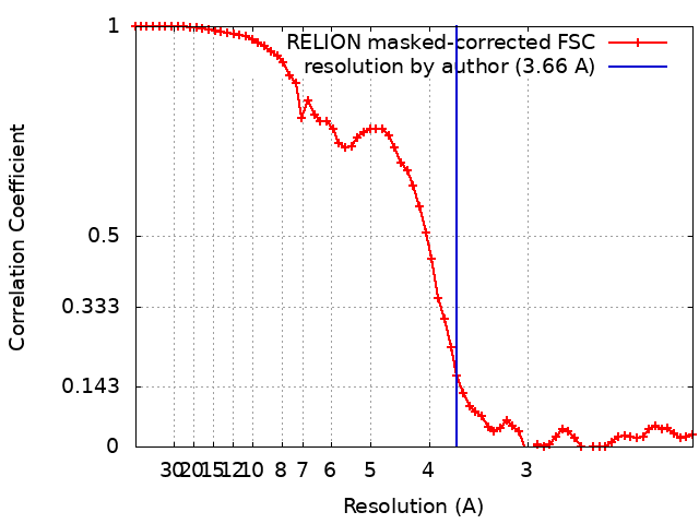
| Method | single particle reconstruction / cryo EM / Resolution: 3.66 Å | |||||||||
 Authors Authors | Martinez Fiesco JA / Ping Z | |||||||||
| Funding support |  United States, 2 items United States, 2 items
| |||||||||
 Citation Citation | Journal: Nat Commun / Year: 2022 Title: Structural insights into the BRAF monomer-to-dimer transition mediated by RAS binding. Authors: Juliana A Martinez Fiesco / David E Durrant / Deborah K Morrison / Ping Zhang / Abstract: RAF kinases are essential effectors of RAS, but how RAS binding initiates the conformational changes needed for autoinhibited RAF monomers to form active dimers has remained unclear. Here, we present ...RAF kinases are essential effectors of RAS, but how RAS binding initiates the conformational changes needed for autoinhibited RAF monomers to form active dimers has remained unclear. Here, we present cryo-electron microscopy structures of full-length BRAF complexes derived from mammalian cells: autoinhibited, monomeric BRAF:14-3-3:MEK and BRAF:14-3-3 complexes, and an inhibitor-bound, dimeric BRAF:14-3-3 complex, at 3.7, 4.1, and 3.9 Å resolution, respectively. In both autoinhibited, monomeric structures, the RAS binding domain (RBD) of BRAF is resolved, revealing that the RBD forms an extensive contact interface with the 14-3-3 protomer bound to the BRAF C-terminal site and that key basic residues required for RBD-RAS binding are exposed. Moreover, through structure-guided mutational studies, our findings indicate that RAS-RAF binding is a dynamic process and that RBD residues at the center of the RBD:14-3-3 interface have a dual function, first contributing to RAF autoinhibition and then to the full spectrum of RAS-RBD interactions. | |||||||||
| History |
|
- Structure visualization
| Movie |
Movie viewer |
|---|---|
| Structure viewer | EM map: SurfViewMolmilJmol/JSmol |
| Supplemental images |
- Downloads & links
-EMDB archive
| Map data | emd_23813.map.gz | 2.6 MB | EMDB map data format | |
|---|---|---|---|---|
| Header (meta data) | emd-23813-v30.xmlemd-23813.xml | 18.7 KB 18.7 KB | Display Display | EMDB header |
| FSC (resolution estimation) | emd_23813_fsc.xml | 6.5 KB | Display | FSC data file |
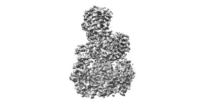
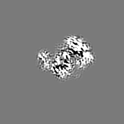
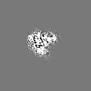
| Images |  emd_23813.png emd_23813.png | 52 KB | ||
| Filedesc metadata | emd-23813.cif.gz | 7.4 KB | ||
| Archive directory |  http://ftp.pdbj.org/pub/emdb/structures/EMD-23813ftp://ftp.pdbj.org/pub/emdb/structures/EMD-23813 http://ftp.pdbj.org/pub/emdb/structures/EMD-23813ftp://ftp.pdbj.org/pub/emdb/structures/EMD-23813 | HTTPS FTP |
-Related structure data
| Related structure data |  7mfdMC  7mfeC 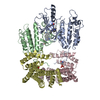 7mffC M: atomic model generated by this map C: citing same article ( |
|---|---|
| Similar structure data |

-Links
| EMDB pages | EMDB (EBI/PDBe) / EMDataResource |
|---|---|
| Related items in Molecule of the Month |


-Map
| File | Download / File: emd_23813.map.gz / Format: CCP4 / Size: 22.2 MB / Type: IMAGE STORED AS FLOATING POINT NUMBER (4 BYTES) | ||||||||||||||||||||||||||||||||||||||||||||||||||||||||||||
|---|---|---|---|---|---|---|---|---|---|---|---|---|---|---|---|---|---|---|---|---|---|---|---|---|---|---|---|---|---|---|---|---|---|---|---|---|---|---|---|---|---|---|---|---|---|---|---|---|---|---|---|---|---|---|---|---|---|---|---|---|---|
| Projections & slices | Image control
Images are generated by Spider. | ||||||||||||||||||||||||||||||||||||||||||||||||||||||||||||
| Voxel size | X=Y=Z: 1.058 Å | ||||||||||||||||||||||||||||||||||||||||||||||||||||||||||||
| Density |
| ||||||||||||||||||||||||||||||||||||||||||||||||||||||||||||
| Symmetry | Space group: 1 | ||||||||||||||||||||||||||||||||||||||||||||||||||||||||||||
| Details | EMDB XML:
CCP4 map header:
| ||||||||||||||||||||||||||||||||||||||||||||||||||||||||||||
 Z (Sec.)
 Y (Row.)
 X (Col.)


-Supplemental data
- Sample components
-Entire : Autoinhibited B-Raf:(14-3-3)2:MEK complex with resolved RBD
| Entire | Name: Autoinhibited B-Raf:(14-3-3)2:MEK complex with resolved RBD |
|---|---|
| Components |
|
-Supramolecule #1: Autoinhibited B-Raf:(14-3-3)2:MEK complex with resolved RBD
| Supramolecule | Name: Autoinhibited B-Raf:(14-3-3)2:MEK complex with resolved RBD type: complex / ID: 1 / Parent: 0 / Macromolecule list: #1-#3 |
|---|
-Supramolecule #2: B-raf
| Supramolecule | Name: B-raf / type: complex / ID: 2 / Parent: 1 / Macromolecule list: #1 |
|---|---|
| Source (natural) | Organism: Homo sapiens (human) |
-Supramolecule #3: MEK
| Supramolecule | Name: MEK / type: complex / ID: 3 / Parent: 1 / Macromolecule list: #2 |
|---|---|
| Source (natural) | Organism: Homo sapiens (human) |
-Supramolecule #4: 14-3-3 protein zeta/delta
| Supramolecule | Name: 14-3-3 protein zeta/delta / type: complex / ID: 4 / Parent: 1 / Macromolecule list: #3 |
|---|---|
| Source (natural) | Organism: Homo sapiens (human) |
-Macromolecule #1: Serine/threonine-protein kinase B-raf
| Macromolecule | Name: Serine/threonine-protein kinase B-raf / type: protein_or_peptide / ID: 1 / Number of copies: 1 / Enantiomer: LEVO / EC number: non-specific serine/threonine protein kinase |
|---|---|
| Source (natural) | Organism: Homo sapiens (human) |
| Molecular weight | Theoretical: 84.697695 KDa |
| Recombinant expression | Organism: Homo sapiens (human) |
| Sequence | String: MAALSGGGGG GAEPGQALFN GDMEPEAGAG AGAAASSAAD PAIPEEVWNI KQMIKLTQEH IEALLDKFGG EHNPPSIYLE AYEEYTSKL DALQQREQQL LESLGNGTDF SVSSSASMDT VTSSSSSSLS VLPSSLSVFQ NPTDVARSNP KSPQKPIVRV F LPNKQRTV ...String: MAALSGGGGG GAEPGQALFN GDMEPEAGAG AGAAASSAAD PAIPEEVWNI KQMIKLTQEH IEALLDKFGG EHNPPSIYLE AYEEYTSKL DALQQREQQL LESLGNGTDF SVSSSASMDT VTSSSSSSLS VLPSSLSVFQ NPTDVARSNP KSPQKPIVRV F LPNKQRTV VPARCGVTVR DSLKKALMMR GLIPECCAVY RIQDGEKKPI GWDTDISWLT GEELHVEVLE NVPLTTHNFV RK TFFTLAF CDFCRKLLFQ GFRCQTCGYK FHQRCSTEVP LMCVNYDQLD LLFVSKFFEH HPIPQEEASL AETALTSGSS PSA PASDSI GPQILTSPSP SKSIPIPQPF RPADEDHRNQ FGQRDRSS(SEP)A PNVHINTIEP VNIDDLIRDQ GFRGDGGSTT GLSATPPAS LPGSLTNVKA LQKSPGPQRE RKSSSSSEDR NRMKTLGRRD SSDDWEIPDG QITVGQRIGS GSFGTVYKGK W HGDVAVKM LNVTAPTPQQ LQAFKNEVGV LRKTRHVNIL LFMGYSTKPQ LAIVTQWCEG SSLYHHLHII ETKFEMIKLI DI ARQTAQG MDYLHAKSII HRDLKSNNIF LHEDLTVKIG DFGLATVKSR WSGSHQFEQL SGSILWMAPE VIRMQDKNPY SFQ SDVYAF GIVLYELMTG QLPYSNINNR DQIIFMVGRG YLSPDLSKVR SNCPKAMKRL MAECLKKKRD ERPLFPQILA SIEL LARSL PKIHRSA(SEP)EP SLNRAGFQTE DFSLYACASP KTPIQAGGYG AFPVH UniProtKB: Serine/threonine-protein kinase B-raf |
-Macromolecule #2: Dual specificity mitogen-activated protein kinase kinase 1
| Macromolecule | Name: Dual specificity mitogen-activated protein kinase kinase 1 type: protein_or_peptide / ID: 2 / Number of copies: 1 / Enantiomer: LEVO / EC number: mitogen-activated protein kinase kinase |
|---|---|
| Source (natural) | Organism: Homo sapiens (human) |
| Molecular weight | Theoretical: 43.493938 KDa |
| Sequence | String: MPKKKPTPIQ LNPAPDGSAV NGTSSAETNL EALQKKLEEL ELDEQQRKRL EAFLTQKQKV GELKDDDFEK ISELGAGNGG VVFKVSHKP SGLVMARKLI HLEIKPAIRN QIIRELQVLH ECNSPYIVGF YGAFYSDGEI SICMEHMDGG SLDQVLKKAG R IPEQILGK ...String: MPKKKPTPIQ LNPAPDGSAV NGTSSAETNL EALQKKLEEL ELDEQQRKRL EAFLTQKQKV GELKDDDFEK ISELGAGNGG VVFKVSHKP SGLVMARKLI HLEIKPAIRN QIIRELQVLH ECNSPYIVGF YGAFYSDGEI SICMEHMDGG SLDQVLKKAG R IPEQILGK VSIAVIKGLT YLREKHKIMH RDVKPSNILV NSRGEIKLCD FGVSGQLIDS MANSFVGTRS YMSPERLQGT HY SVQSDIW SMGLSLVEMA VGRYPIPPPD AKELELMFGC QVEGDAAETP PRPRTPGRPL SSYGMDSRPP MAIFELLDYI VNE PPPKLP SGVFSLEFQD FVNKCLIKNP AERADLKQLM VHAFIKRSDA EEVDFAGWLC STIGLNQPST PTHAAGV UniProtKB: Dual specificity mitogen-activated protein kinase kinase 1 |
-Macromolecule #3: 14-3-3 protein zeta/delta
| Macromolecule | Name: 14-3-3 protein zeta/delta / type: protein_or_peptide / ID: 3 / Number of copies: 2 / Enantiomer: LEVO |
|---|---|
| Source (natural) | Organism: Homo sapiens (human) |
| Molecular weight | Theoretical: 27.777092 KDa |
| Sequence | String: MDKNELVQKA KLAEQAERYD DMAACMKSVT EQGAELSNEE RNLLSVAYKN VVGARRSSWR VVSSIEQKTE GAEKKQQMAR EYREKIETE LRDICNDVLS LLEKFLIPNA SQAESKVFYL KMKGDYYRYL AEVAAGDDKK GIVDQSQQAY QEAFEISKKE M QPTHPIRL ...String: MDKNELVQKA KLAEQAERYD DMAACMKSVT EQGAELSNEE RNLLSVAYKN VVGARRSSWR VVSSIEQKTE GAEKKQQMAR EYREKIETE LRDICNDVLS LLEKFLIPNA SQAESKVFYL KMKGDYYRYL AEVAAGDDKK GIVDQSQQAY QEAFEISKKE M QPTHPIRL GLALNFSVFY YEILNSPEKA CSLAKTAFDE AIAELDTLSE ESYKDSTLIM QLLRDNLTLW TSDTQGDEAE AG EGGEN UniProtKB: 14-3-3 protein zeta/delta |
-Macromolecule #4: ZINC ION
| Macromolecule | Name: ZINC ION / type: ligand / ID: 4 / Number of copies: 2 / Formula: ZN |
|---|---|
| Molecular weight | Theoretical: 65.409 Da |
-Macromolecule #5: N-(3-fluoro-4-{[4-methyl-2-oxo-7-(pyrimidin-2-yloxy)-2H-chromen-3...
| Macromolecule | Name: N-(3-fluoro-4-{[4-methyl-2-oxo-7-(pyrimidin-2-yloxy)-2H-chromen-3-yl]methyl}pyridin-2-yl)-N'-methylsulfuric diamide type: ligand / ID: 5 / Number of copies: 1 / Formula: CHU |
|---|---|
| Molecular weight | Theoretical: 471.462 Da |
| Chemical component information |  ChemComp-CHU: |
-Experimental details
-Structure determination
| Method | cryo EM |
|---|---|
 Processing Processing | single particle reconstruction |
| Aggregation state | particle |
-Sample preparation
| Concentration | 0.2 mg/mL | ||||||||||||
|---|---|---|---|---|---|---|---|---|---|---|---|---|---|
| Buffer | pH: 8 Component:
| ||||||||||||
| Grid | Model: Quantifoil R1.2/1.3 / Material: GOLD / Mesh: 300 / Pretreatment - Type: GLOW DISCHARGE / Pretreatment - Time: 45 sec. / Details: 20mAmp | ||||||||||||
| Vitrification | Cryogen name: ETHANE / Chamber humidity: 90 % / Chamber temperature: 277.15 K / Instrument: FEI VITROBOT MARK IV |
- Electron microscopy
| Microscope | FEI TITAN KRIOS |
|---|---|
| Image recording | Film or detector model: GATAN K2 SUMMIT (4k x 4k) / Detector mode: SUPER-RESOLUTION / Average electron dose: 57.0 e/Å2 |
| Electron beam | Acceleration voltage: 300 kV / Electron source:  FIELD EMISSION GUN FIELD EMISSION GUN |
| Electron optics | Illumination mode: FLOOD BEAM / Imaging mode: BRIGHT FIELD |
| Experimental equipment |  Model: Titan Krios / Image courtesy: FEI Company |