National Institutes of Health/National Institute Of Allergy and Infectious Diseases (NIH/NIAID)
75N93019C00051
United States
Citation
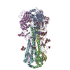
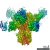
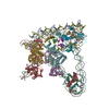
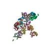
Journal: Proc Natl Acad Sci U S A / Year: 2021 Title: Influenza hemagglutinin-specific IgA Fc-effector functionality is restricted to stalk epitopes. Authors: Alec W Freyn / Julianna Han / Jenna J Guthmiller / Mark J Bailey / Karlynn Neu / Hannah L Turner / Victoria C Rosado / Veronika Chromikova / Min Huang / Shirin Strohmeier / Sean T H Liu / ...Authors: Alec W Freyn / Julianna Han / Jenna J Guthmiller / Mark J Bailey / Karlynn Neu / Hannah L Turner / Victoria C Rosado / Veronika Chromikova / Min Huang / Shirin Strohmeier / Sean T H Liu / Viviana Simon / Florian Krammer / Andrew B Ward / Peter Palese / Patrick C Wilson / Raffael Nachbagauer / Abstract: In this study, we utilized a panel of human immunoglobulin (Ig) IgA monoclonal antibodies isolated from the plasmablasts of eight donors after 2014/2015 influenza virus vaccination (Fluarix) to study ...In this study, we utilized a panel of human immunoglobulin (Ig) IgA monoclonal antibodies isolated from the plasmablasts of eight donors after 2014/2015 influenza virus vaccination (Fluarix) to study the binding and functional specificities of this isotype. In this cohort, isolated IgA monoclonal antibodies were primarily elicited against the hemagglutinin protein of the H1N1 component of the vaccine. To compare effector functionalities, an H1-specific subset of antibodies targeting distinct epitopes were expressed as monomeric, dimeric, or secretory IgA, as well as in an IgG1 backbone. When expressed with an IgG Fc domain, all antibodies elicited Fc-effector activity in a primary polymorphonuclear cell-based assay which differs from previous observations that found only stalk-specific antibodies activate the low-affinity FcγRIIIa. However, when expressed with IgA Fc domains, only antibodies targeting the stalk domain showed Fc-effector activity in line with these previous findings. To identify the cause of this discrepancy, we then confirmed that IgG signaling through the high-affinity FcγI receptor was not restricted to stalk epitopes. Since no corresponding high-affinity Fcα receptor exists, the IgA repertoire may therefore be limited to stalk-specific epitopes in the context of Fc receptor signaling.
History
Deposition
Jan 20, 2021
-
Header (metadata) release
Mar 3, 2021
-
Map release
Mar 3, 2021
-
Update
Mar 3, 2021
-
Current status
Mar 3, 2021
Processing site: RCSB / Status: Released
-
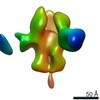
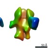
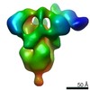
Structure visualization
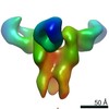
Movie
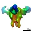
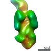
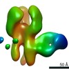
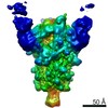
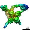
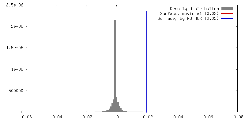
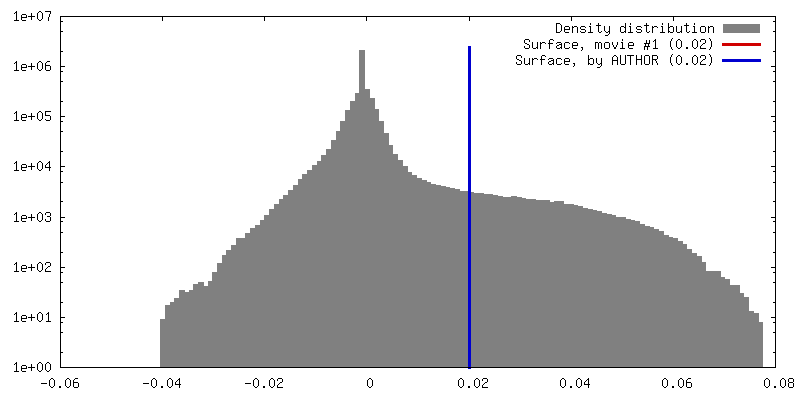
Surface view with section colored by density value
In the structure databanks used in Yorodumi, some data are registered as the other names, "COVID-19 virus" and "2019-nCoV". Here are the details of the virus and the list of structure data.
Jan 31, 2019. EMDB accession codes are about to change! (news from PDBe EMDB page)
EMDB accession codes are about to change! (news from PDBe EMDB page)
The allocation of 4 digits for EMDB accession codes will soon come to an end. Whilst these codes will remain in use, new EMDB accession codes will include an additional digit and will expand incrementally as the available range of codes is exhausted. The current 4-digit format prefixed with “EMD-” (i.e. EMD-XXXX) will advance to a 5-digit format (i.e. EMD-XXXXX), and so on. It is currently estimated that the 4-digit codes will be depleted around Spring 2019, at which point the 5-digit format will come into force.
The EM Navigator/Yorodumi systems omit the EMD- prefix.
Related info.:Q: What is EMD? / ID/Accession-code notation in Yorodumi/EM Navigator
Yorodumi is a browser for structure data from EMDB, PDB, SASBDB, etc.
This page is also the successor to EM Navigator detail page, and also detail information page/front-end page for Omokage search.
The word "yorodu" (or yorozu) is an old Japanese word meaning "ten thousand". "mi" (miru) is to see.
Related info.:EMDB / PDB / SASBDB / Comparison of 3 databanks / Yorodumi Search / Aug 31, 2016. New EM Navigator & Yorodumi / Yorodumi Papers / Jmol/JSmol / Function and homology information / Changes in new EM Navigator and Yorodumi
 Movie
Movie Controller

 Yorodumi
 Open data
 Basic information
 Map data
 Sample
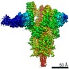
 Homo sapiens (human) /
 Influenza A virus
 Authors
 United States, 1 items
  Citation
 Structure visualization
 Movie viewer
 Downloads & links
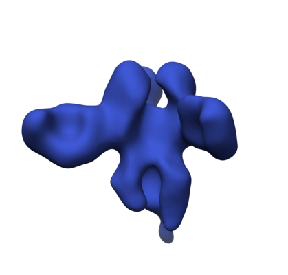 emd_23314.png
 http://ftp.pdbj.org/pub/emdb/structures/EMD-23314
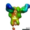
 Z (Sec.)
 Y (Row.)
 X (Col.)


 Sample components
 Processing
 Electron microscopy
 FIELD EMISSION GUN