 Movie
Movie Controller

[English] 日本語
 Yorodumi
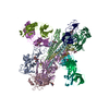
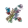
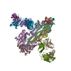
- PDB-5uqy: Crystal structure of Marburg virus GP in complex with the human s... -
+ Open data

- Basic information
| Entry | Database: PDB / ID: 5uqy | ||||||||||||
|---|---|---|---|---|---|---|---|---|---|---|---|---|---|
| Title | Crystal structure of Marburg virus GP in complex with the human survivor antibody MR78 | ||||||||||||
 Components Components |
| ||||||||||||
 Keywords Keywords | VIRAL PROTEIN/IMMUNE SYSTEM / glycoprotein / viral protein / antibody / Fab / VIRAL PROTEIN-IMMUNE SYSTEM complex | ||||||||||||
| Function / homology |  Function and homology information Function and homology informationfusion of virus membrane with host endosome membrane / viral envelope / symbiont entry into host cell / virion attachment to host cell / host cell plasma membrane / virion membrane Similarity search - Function | ||||||||||||
| Biological species |  Lake Victoria marburgvirus Lake Victoria marburgvirus Homo sapiens (human) Homo sapiens (human) | ||||||||||||
| Method |  X-RAY DIFFRACTION / SYNCHROTRON / MOLECULAR REPLACEMENT / Resolution: 3.6 Å X-RAY DIFFRACTION / SYNCHROTRON / MOLECULAR REPLACEMENT / Resolution: 3.6 Å | ||||||||||||
 Authors Authors | Hashiguchi, T. / Fusco, M.L. / Hastie, K.M. / Bomholdt, Z.A. / Lee, J.E. / Flyak, A.I. / Matsuoka, R. / Kohda, D. / Yanagi, Y. / Hammel, M. ...Hashiguchi, T. / Fusco, M.L. / Hastie, K.M. / Bomholdt, Z.A. / Lee, J.E. / Flyak, A.I. / Matsuoka, R. / Kohda, D. / Yanagi, Y. / Hammel, M. / Crowe, J.E. / Saphire, E.O. | ||||||||||||
| Funding support |  United States, 3items United States, 3items
| ||||||||||||
 Citation Citation | Journal: Cell / Year: 2015 Title: Structural basis for Marburg virus neutralization by a cross-reactive human antibody. Authors: Hashiguchi, T. / Fusco, M.L. / Bornholdt, Z.A. / Lee, J.E. / Flyak, A.I. / Matsuoka, R. / Kohda, D. / Yanagi, Y. / Hammel, M. / Crowe, J.E. / Saphire, E.O. | ||||||||||||
| History |
|
- Structure visualization
| Structure viewer | Molecule: MolmilJmol/JSmol |
|---|
- Downloads & links
-Download
| PDBx/mmCIF format | 5uqy.cif.gz | 557.7 KB | Display | PDBx/mmCIF format |
|---|---|---|---|---|
| PDB format | pdb5uqy.ent.gz | 440.2 KB | Display | PDB format |
| PDBx/mmJSON format | 5uqy.json.gz | Tree view | PDBx/mmJSON format | |
| Others |  Other downloads Other downloads |
-Validation report
| Arichive directory | https://data.pdbj.org/pub/pdb/validation_reports/uq/5uqyftp://data.pdbj.org/pub/pdb/validation_reports/uq/5uqy | HTTPS FTP |
|---|
-Related structure data
| Related structure data | 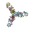 3csyS S: Starting model for refinement |
|---|---|
| Similar structure data |


-Links
 PDBj
PDBj


- Assembly
| Deposited unit | 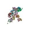
| ||||||||
|---|---|---|---|---|---|---|---|---|---|
| 1 | 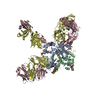
| ||||||||
| 2 | 
| ||||||||
| Unit cell |
|
-Components
-ENVELOPE GLYCOPROTEIN ... , 2 types, 8 molecules AEIMBFJN
| #1: Protein | Mass: 27885.434 Da / Num. of mol.: 4 / Fragment: UNP residues 17-256, 426-435 Source method: isolated from a genetically manipulated source Source: (gene. exp.) Lake Victoria marburgvirus (strain Ravn-87)Strain: Ravn-87 / Gene: GP / Plasmid: pMT / Cell line (production host): S2 / Production host:  #2: Protein | Mass: 26137.631 Da / Num. of mol.: 4 / Fragment: UNP residues 436-637 Source method: isolated from a genetically manipulated source Source: (gene. exp.) Lake Victoria marburgvirus (strain Ravn-87)Strain: Ravn-87 / Gene: GP / Plasmid: pMT / Cell line (production host): S2 / Production host: |
|---|
-Antibody , 2 types, 8 molecules CGKODHLP
| #3: Antibody | Mass: 23307.877 Da / Num. of mol.: 4 / Source method: isolated from a natural source / Source: (natural) Homo sapiens (human) / Cell: Hybridoma / Plasmid details: B cell hybrdoma#4: Antibody | Mass: 23869.578 Da / Num. of mol.: 4 / Source method: isolated from a natural source / Source: (natural) Homo sapiens (human) / Cell: Hybridoma / Plasmid details: B cell hybrdoma |
|---|
-Sugars , 6 types, 14 molecules 

| #5: Polysaccharide | 2-acetamido-2-deoxy-beta-D-glucopyranose-(1-4)-2-acetamido-2-deoxy-beta-D-glucopyranose Source method: isolated from a genetically manipulated source #6: Polysaccharide | Source method: isolated from a genetically manipulated source #7: Polysaccharide | Source method: isolated from a genetically manipulated source #8: Polysaccharide | alpha-D-mannopyranose-(1-2)-alpha-D-mannopyranose-(1-3)-beta-D-mannopyranose-(1-4)-2-acetamido-2- ...alpha-D-mannopyranose-(1-2)-alpha-D-mannopyranose-(1-3)-beta-D-mannopyranose-(1-4)-2-acetamido-2-deoxy-beta-D-glucopyranose-(1-4)-2-acetamido-2-deoxy-beta-D-glucopyranose | Source method: isolated from a genetically manipulated source #9: Sugar | ChemComp-NAG / |  Type: D-saccharide, beta linking / Mass: 221.208 Da / Num. of mol.: 1 Type: D-saccharide, beta linking / Mass: 221.208 Da / Num. of mol.: 1Source method: isolated from a genetically manipulated source Formula: C8H15NO6 #10: Sugar |  Type: D-saccharide, alpha linking / Mass: 180.156 Da / Num. of mol.: 2 Type: D-saccharide, alpha linking / Mass: 180.156 Da / Num. of mol.: 2Source method: isolated from a genetically manipulated source Formula: C6H12O6 |
|---|
-Details
| Has protein modification | Y |
|---|
-Experimental details
-Experiment
| Experiment | Method: X-RAY DIFFRACTION / Number of used crystals: 1 |
|---|
- Sample preparation
| Crystal | Density Matthews: 2.88 Å3/Da / Density % sol: 57.32 % |
|---|---|
| Crystal grow | Temperature: 293 K / Method: vapor diffusion, hanging drop / pH: 6.5 Details: 0.1 M NaCl, 0.05 M MES pH6.5, 13 % PEG4000, 0.5 % ethyl acetate |
-Data collection
| Diffraction | Mean temperature: 100 K |
|---|---|
| Diffraction source | Source: SYNCHROTRON / Site: Photon Factory  / Beamline: BL-1A / Wavelength: 1.1 Å / Beamline: BL-1A / Wavelength: 1.1 Å |
| Detector | Type: DECTRIS PILATUS 2M-F / Detector: PIXEL / Date: Feb 2, 2014 |
| Radiation | Protocol: SINGLE WAVELENGTH / Monochromatic (M) / Laue (L): M / Scattering type: x-ray |
| Radiation wavelength | Wavelength: 1.1 Å / Relative weight: 1 |
| Reflection | Resolution: 3.6→130.53 Å / Num. obs: 52806 / % possible obs: 99.4 % / Redundancy: 5.1 % / CC1/2: 0.994 / Rsym value: 0.144 / Net I/σ(I): 12.9 |
| Reflection shell | Resolution: 3.6→3.612 Å / Redundancy: 5.1 % / Mean I/σ(I) obs: 2.1 / Num. unique obs: 534 / Rsym value: 0.94 / % possible all: 100 |
- Processing
| Software |
| ||||||||||||||||||||||||||||||||||||||||||||||||||||||||||||||||||||||||||||||||||||||||||||||||||||||||||||||||||||||||||||||||||||||||||||
|---|---|---|---|---|---|---|---|---|---|---|---|---|---|---|---|---|---|---|---|---|---|---|---|---|---|---|---|---|---|---|---|---|---|---|---|---|---|---|---|---|---|---|---|---|---|---|---|---|---|---|---|---|---|---|---|---|---|---|---|---|---|---|---|---|---|---|---|---|---|---|---|---|---|---|---|---|---|---|---|---|---|---|---|---|---|---|---|---|---|---|---|---|---|---|---|---|---|---|---|---|---|---|---|---|---|---|---|---|---|---|---|---|---|---|---|---|---|---|---|---|---|---|---|---|---|---|---|---|---|---|---|---|---|---|---|---|---|---|---|---|---|
| Refinement | Method to determine structure: MOLECULAR REPLACEMENT Starting model: 3CSY Resolution: 3.6→84.739 Å / SU ML: 0.48 / Cross valid method: FREE R-VALUE / σ(F): 1.33 / Phase error: 26.52
| ||||||||||||||||||||||||||||||||||||||||||||||||||||||||||||||||||||||||||||||||||||||||||||||||||||||||||||||||||||||||||||||||||||||||||||
| Solvent computation | Shrinkage radii: 0.9 Å / VDW probe radii: 1.11 Å | ||||||||||||||||||||||||||||||||||||||||||||||||||||||||||||||||||||||||||||||||||||||||||||||||||||||||||||||||||||||||||||||||||||||||||||
| Refinement step | Cycle: LAST / Resolution: 3.6→84.739 Å
| ||||||||||||||||||||||||||||||||||||||||||||||||||||||||||||||||||||||||||||||||||||||||||||||||||||||||||||||||||||||||||||||||||||||||||||
| Refine LS restraints |
| ||||||||||||||||||||||||||||||||||||||||||||||||||||||||||||||||||||||||||||||||||||||||||||||||||||||||||||||||||||||||||||||||||||||||||||
| LS refinement shell |
|
