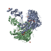
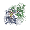
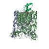
+ Open data
- Basic information
| Entry | Database: EMDB / ID: EMD-22697 | |||||||||
|---|---|---|---|---|---|---|---|---|---|---|
| Title | CBF3 | |||||||||
 Map data Map data | CBF3 | |||||||||
 Sample Sample |
| |||||||||
 Keywords Keywords | CBF3 / DNA BINDING PROTEIN | |||||||||
| Function / homology |  Function and homology information Function and homology informationRAVE complex / Iron uptake and transport / CBF3 complex / regulation of transcription by galactose / : / cellular response to methylmercury / vacuolar proton-transporting V-type ATPase complex assembly / septin ring assembly / centromeric DNA binding / regulation of exit from mitosis ...RAVE complex / Iron uptake and transport / CBF3 complex / regulation of transcription by galactose / : / cellular response to methylmercury / vacuolar proton-transporting V-type ATPase complex assembly / septin ring assembly / centromeric DNA binding / regulation of exit from mitosis / kinetochore assembly / exit from mitosis / positive regulation of D-glucose transmembrane transport / vacuolar acidification / protein neddylation / mitotic intra-S DNA damage checkpoint signaling / mitochondrial fusion / silent mating-type cassette heterochromatin formation / DNA binding, bending / regulation of metabolic process / SCF ubiquitin ligase complex / SCF-dependent proteasomal ubiquitin-dependent protein catabolic process / FBXL7 down-regulates AURKA during mitotic entry and in early mitosis / mitotic spindle assembly checkpoint signaling / Orc1 removal from chromatin / cullin family protein binding / Antigen processing: Ubiquitination & Proteasome degradation / DNA replication origin binding / regulation of protein-containing complex assembly / subtelomeric heterochromatin formation / negative regulation of cytoplasmic translation / endomembrane system / regulation of mitotic cell cycle / G1/S transition of mitotic cell cycle / kinetochore / G2/M transition of mitotic cell cycle / mitotic cell cycle / protein-containing complex assembly / ubiquitin-dependent protein catabolic process / DNA-binding transcription factor activity, RNA polymerase II-specific / chromosome, telomeric region / protein ubiquitination / zinc ion binding / identical protein binding / nucleus / cytoplasm Similarity search - Function | |||||||||
| Biological species |   | |||||||||
| Method | single particle reconstruction / cryo EM / Resolution: 4.0 Å | |||||||||
 Authors Authors | Ruifang G / Yawen B | |||||||||
 Citation Citation |  Journal: Nat Commun / Year: 2021 Journal: Nat Commun / Year: 2021Title: Structural and dynamic mechanisms of CBF3-guided centromeric nucleosome formation. Authors: Ruifang Guan / Tengfei Lian / Bing-Rui Zhou / Emily He / Carl Wu / Martin Singleton / Yawen Bai /   Abstract: Accurate chromosome segregation relies on the specific centromeric nucleosome-kinetochore interface. In budding yeast, the centromere CBF3 complex guides the deposition of CENP-A, an H3 variant, to ...Accurate chromosome segregation relies on the specific centromeric nucleosome-kinetochore interface. In budding yeast, the centromere CBF3 complex guides the deposition of CENP-A, an H3 variant, to form the centromeric nucleosome in a DNA sequence-dependent manner. Here, we determine the structures of the centromeric nucleosome containing the native CEN3 DNA and the CBF3core bound to the canonical nucleosome containing an engineered CEN3 DNA. The centromeric nucleosome core structure contains 115 base pair DNA including a CCG motif. The CBF3core specifically recognizes the nucleosomal CCG motif through the Gal4 domain while allosterically altering the DNA conformation. Cryo-EM, modeling, and mutational studies reveal that the CBF3core forms dynamic interactions with core histones H2B and CENP-A in the CEN3 nucleosome. Our results provide insights into the structure of the budding yeast centromeric nucleosome and the mechanism of its assembly, which have implications for analogous processes of human centromeric nucleosome formation. | |||||||||
| History |
|
- Structure visualization
| Movie |
 Movie viewer Movie viewer |
|---|---|
| Structure viewer | EM map:  SurfView SurfView Molmil Molmil Jmol/JSmol Jmol/JSmol |
| Supplemental images |
- Downloads & links
-EMDB archive
| Map data |  emd_22697.map.gz emd_22697.map.gz | 78.4 MB |  EMDB map data format EMDB map data format | |
|---|---|---|---|---|
| Header (meta data) |  emd-22697-v30.xml emd-22697-v30.xml emd-22697.xml emd-22697.xml | 11.8 KB 11.8 KB | Display Display |  EMDB header EMDB header |
| Images |  emd_22697.png emd_22697.png | 161.4 KB | ||
| Filedesc metadata |  emd-22697.cif.gz emd-22697.cif.gz | 5.7 KB | ||
| Archive directory |  http://ftp.pdbj.org/pub/emdb/structures/EMD-22697 http://ftp.pdbj.org/pub/emdb/structures/EMD-22697 ftp://ftp.pdbj.org/pub/emdb/structures/EMD-22697 ftp://ftp.pdbj.org/pub/emdb/structures/EMD-22697 | HTTPS FTP |
-Validation report
| Summary document |  emd_22697_validation.pdf.gz emd_22697_validation.pdf.gz | 501.9 KB | Display |  EMDB validaton report EMDB validaton report |
|---|---|---|---|---|
| Full document |  emd_22697_full_validation.pdf.gz emd_22697_full_validation.pdf.gz | 501.5 KB | Display | |
| Data in XML |  emd_22697_validation.xml.gz emd_22697_validation.xml.gz | 6.2 KB | Display | |
| Data in CIF |  emd_22697_validation.cif.gz emd_22697_validation.cif.gz | 7.1 KB | Display | |
| Arichive directory |  https://ftp.pdbj.org/pub/emdb/validation_reports/EMD-22697 https://ftp.pdbj.org/pub/emdb/validation_reports/EMD-22697 ftp://ftp.pdbj.org/pub/emdb/validation_reports/EMD-22697 ftp://ftp.pdbj.org/pub/emdb/validation_reports/EMD-22697 | HTTPS FTP |
-Related structure data
| Related structure data |  7k79MC  7k78C  7k7gC M: atomic model generated by this map C: citing same article ( |
|---|---|
| Similar structure data |
- Links
Links
| EMDB pages |  EMDB (EBI/PDBe) / EMDB (EBI/PDBe) /  EMDataResource EMDataResource |
|---|---|
| Related items in Molecule of the Month |
- Map
Map
| File |  Download / File: emd_22697.map.gz / Format: CCP4 / Size: 83.7 MB / Type: IMAGE STORED AS FLOATING POINT NUMBER (4 BYTES) Download / File: emd_22697.map.gz / Format: CCP4 / Size: 83.7 MB / Type: IMAGE STORED AS FLOATING POINT NUMBER (4 BYTES) | ||||||||||||||||||||||||||||||||||||||||||||||||||||||||||||||||||||
|---|---|---|---|---|---|---|---|---|---|---|---|---|---|---|---|---|---|---|---|---|---|---|---|---|---|---|---|---|---|---|---|---|---|---|---|---|---|---|---|---|---|---|---|---|---|---|---|---|---|---|---|---|---|---|---|---|---|---|---|---|---|---|---|---|---|---|---|---|---|
| Annotation | CBF3 | ||||||||||||||||||||||||||||||||||||||||||||||||||||||||||||||||||||
| Projections & slices | Image control
Images are generated by Spider. | ||||||||||||||||||||||||||||||||||||||||||||||||||||||||||||||||||||
| Voxel size | X=Y=Z: 1.06 Å | ||||||||||||||||||||||||||||||||||||||||||||||||||||||||||||||||||||
| Density |
| ||||||||||||||||||||||||||||||||||||||||||||||||||||||||||||||||||||
| Symmetry | Space group: 1 | ||||||||||||||||||||||||||||||||||||||||||||||||||||||||||||||||||||
| Details | EMDB XML:
CCP4 map header:
| ||||||||||||||||||||||||||||||||||||||||||||||||||||||||||||||||||||
-Supplemental data
- Sample components
-Entire : CBF3CORE
| Entire | Name: CBF3CORE |
|---|---|
| Components |
|
-Supramolecule #1: CBF3CORE
| Supramolecule | Name: CBF3CORE / type: complex / ID: 1 / Parent: 0 / Macromolecule list: #1-#2 |
|---|---|
| Source (natural) | Organism:  |
-Macromolecule #1: Centromere DNA-binding protein complex CBF3 subunit C
| Macromolecule | Name: Centromere DNA-binding protein complex CBF3 subunit C / type: protein_or_peptide / ID: 1 / Number of copies: 1 / Enantiomer: LEVO |
|---|---|
| Source (natural) | Organism:  Strain: ATCC 204508 / S288c |
| Molecular weight | Theoretical: 60.899961 KDa |
| Recombinant expression | Organism:  |
| Sequence | String: MGPSFNPVRF LELPIDIRKE VYFHLDGNFC GAHPYPIDIL YKSNDVELPG KPSYKRSKRS KKLLRYMYPV FATYLNIFEY SPQLIEKWL EYAFWLRYDC LVLDCFKVNH LYDGTLIDAL EWTYLDNELR LAYFNKASML EVWYTFKEYK KWVIDSVAFD E LDLLNVSN ...String: MGPSFNPVRF LELPIDIRKE VYFHLDGNFC GAHPYPIDIL YKSNDVELPG KPSYKRSKRS KKLLRYMYPV FATYLNIFEY SPQLIEKWL EYAFWLRYDC LVLDCFKVNH LYDGTLIDAL EWTYLDNELR LAYFNKASML EVWYTFKEYK KWVIDSVAFD E LDLLNVSN IQFNIDNLTP QLVDKCLSIL EQKDLFATIG EVQFGQDEEV GEEKDVDVSG ANSDENSSPS STIKNKKRSA SK RSHSDNG NVGATHNQLT SISVIRTIRS MESMKSLRKI TVRGEKLYEL LINFHGFRDN PGKTISYIVK RRINEIRLSR MNQ ISRTGL ADFTRWDNLQ KLVLSRVAYI DLNSIVFPKN FKSLTMKRVS KIKWWNIEEN ILKELKVDKR TFKSLYIKED DSKF TKFFN LRHTRIKELD KSEINQITYL RCQAIVWLSF RTLNHIKLQN VSEVFNNIIV PRALFDSKRV EIYRCEKISQ VLVIG SRSG SENLYFQGSK RRWKKNFIAV SAANRFKKIS SSGAL UniProtKB: Centromere DNA-binding protein complex CBF3 subunit C |
-Macromolecule #2: Centromere DNA-binding protein complex CBF3 subunit B
| Macromolecule | Name: Centromere DNA-binding protein complex CBF3 subunit B / type: protein_or_peptide / ID: 2 / Number of copies: 2 / Enantiomer: LEVO |
|---|---|
| Source (natural) | Organism:  Strain: ATCC 204508 / S288c |
| Molecular weight | Theoretical: 72.637117 KDa |
| Recombinant expression | Organism:  |
| Sequence | String: MFNRTTQLKS KHPCSVCTRR KVKCDRMIPC GNCRKRGQDS ECMKSTKLIT ASSSKEYLPD LLLFWQNYEY WITNIGLYKT KQRDLTRTP ANLDTDTEEC MFWMNYLQKD QSFQLMNFAM ENLGALYFGS IGDISELYLR VEQYWDRRAD KNHSVDGKYW D ALIWSVFT ...String: MFNRTTQLKS KHPCSVCTRR KVKCDRMIPC GNCRKRGQDS ECMKSTKLIT ASSSKEYLPD LLLFWQNYEY WITNIGLYKT KQRDLTRTP ANLDTDTEEC MFWMNYLQKD QSFQLMNFAM ENLGALYFGS IGDISELYLR VEQYWDRRAD KNHSVDGKYW D ALIWSVFT MCIYYMPVEK LAEIFSVYPL HEYLGSNKRL NWEDGMQLVM CQNFARCSLF QLKQCDFMAH PDIRLVQAYL IL ATTTFPY DEPLLANSLL TQCIHTFKNF HVDDFRPLLN DDPVESIAKV TLGRIFYRLC GCDYLQSGPR KPIALHTEVS SLL QHAAYL QDLPNVDVYR EENSTEVLYW KIISLDRDLD QYLNKSSKPP LKTLDAIRRE LDIFQYKVDS LEEDFRSNNS RFQK FIALF QISTVSWKLF KMYLIYYDTA DSLLKVIHYS KVIISLIVNN FHAKSEFFNR HPMVMQTITR VVSFISFYQI FVESA AVKQ LLVDLTELTA NLPTIFGSKL DKLVYLTERL SKLKLLWDKV QLLDSGDSFY HPVFKILQND IKIIELKNDE MFSLIK GLG SLVPLNKLRQ ESLLEEEDEN NTEPSDFRTI VEEFQSEYNI SDILSGSGGS GENLYFQ UniProtKB: Centromere DNA-binding protein complex CBF3 subunit B |
-Macromolecule #3: Suppressor of kinetochore protein 1
| Macromolecule | Name: Suppressor of kinetochore protein 1 / type: protein_or_peptide / ID: 3 / Number of copies: 1 / Enantiomer: LEVO |
|---|---|
| Source (natural) | Organism:  Strain: ATCC 204508 / S288c |
| Molecular weight | Theoretical: 22.35727 KDa |
| Recombinant expression | Organism:  |
| Sequence | String: MVTSNVVLVS GEGERFTVDK KIAERSLLLK NYLNDMHDSN LQNNSDSESD SDSETNHKSK DNNNGDDDDE DDDEIVMPVP NVRSSVLQK VIEWAEHHRD SNFPDEDDDD SRKSAPVDSW DREFLKVDQE MLYEIILAAN YLNIKPLLDA GCKVVAEMIR G RSPEEIRR ...String: MVTSNVVLVS GEGERFTVDK KIAERSLLLK NYLNDMHDSN LQNNSDSESD SDSETNHKSK DNNNGDDDDE DDDEIVMPVP NVRSSVLQK VIEWAEHHRD SNFPDEDDDD SRKSAPVDSW DREFLKVDQE MLYEIILAAN YLNIKPLLDA GCKVVAEMIR G RSPEEIRR TFNIVNDFTP EEEAAIRREN EWAEDR UniProtKB: Suppressor of kinetochore protein 1 |
-Experimental details
-Structure determination
| Method | cryo EM |
|---|---|
 Processing Processing | single particle reconstruction |
| Aggregation state | particle |
- Sample preparation
| Buffer | pH: 7.3 |
|---|---|
| Vitrification | Cryogen name: ETHANE |
- Electron microscopy
| Microscope | FEI TITAN KRIOS |
|---|---|
| Image recording | Film or detector model: GATAN K2 SUMMIT (4k x 4k) / Average electron dose: 71.0 e/Å2 |
| Electron beam | Acceleration voltage: 300 kV / Electron source:  FIELD EMISSION GUN FIELD EMISSION GUN |
| Electron optics | Illumination mode: SPOT SCAN / Imaging mode: BRIGHT FIELD |
| Experimental equipment |  Model: Titan Krios / Image courtesy: FEI Company |
- Image processing
| Startup model | Type of model: PDB ENTRY |
|---|---|
| Final reconstruction | Resolution.type: BY AUTHOR / Resolution: 4.0 Å / Resolution method: FSC 0.143 CUT-OFF / Number images used: 115666 |
| Initial angle assignment | Type: MAXIMUM LIKELIHOOD |
| Final angle assignment | Type: MAXIMUM LIKELIHOOD |
 Movie
Movie Controller


 Z (Sec.)
 Y (Row.)
 X (Col.)


