 Movie
Movie Controller

[English] 日本語
 Yorodumi
- EMDB-22285: Cryo-EM structure of human ZnT8 double mutant - D110N and D224N, ... -
+ Open data

- Basic information
| Entry | Database: EMDB / ID: EMD-22285 | |||||||||
|---|---|---|---|---|---|---|---|---|---|---|
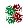
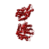
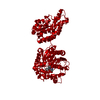
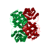
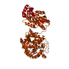
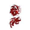
| Title | Cryo-EM structure of human ZnT8 double mutant - D110N and D224N, determined in outward-facing conformation | |||||||||
 Map data Map data | Cryo-EM structure of human ZnT8 with two mutations introduced (D110N and D224N), in the outward-facing conformation | |||||||||
 Sample Sample |
| |||||||||
 Keywords Keywords | ZnT8 / zinc transporter / TRANSPORT PROTEIN | |||||||||
| Function / homology |  Function and homology information Function and homology informationzinc ion import across plasma membrane / insulin processing / zinc:proton antiporter activity / Zinc efflux and compartmentalization by the SLC30 family / zinc ion import into organelle / zinc ion transport / zinc ion transmembrane transporter activity / zinc ion transmembrane transport / regulation of vesicle-mediated transport / intracellular zinc ion homeostasis ...zinc ion import across plasma membrane / insulin processing / zinc:proton antiporter activity / Zinc efflux and compartmentalization by the SLC30 family / zinc ion import into organelle / zinc ion transport / zinc ion transmembrane transporter activity / zinc ion transmembrane transport / regulation of vesicle-mediated transport / intracellular zinc ion homeostasis / insulin secretion / response to zinc ion / Insulin processing / transport vesicle membrane / response to type II interferon / response to glucose / secretory granule / response to interleukin-1 / secretory granule membrane / positive regulation of insulin secretion / cytoplasmic vesicle / Golgi membrane / protein homodimerization activity / zinc ion binding / plasma membrane Similarity search - Function | |||||||||
| Biological species |  Homo sapiens (human) Homo sapiens (human) | |||||||||
| Method | single particle reconstruction / cryo EM / Resolution: 3.8 Å | |||||||||
 Authors Authors | Bai XC / Xue J | |||||||||
 Citation Citation | Journal: Elife / Year: 2020 Title: Cryo-EM structures of human ZnT8 in both outward- and inward-facing conformations. Authors: Jing Xue / Tian Xie / Weizhong Zeng / Youxing Jiang / Xiao-Chen Bai /  Abstract: ZnT8 is a Zn/H antiporter that belongs to SLC30 family and plays an essential role in regulating Zn accumulation in the insulin secretory granules of pancreatic β cells. However, the Zn/H exchange ...ZnT8 is a Zn/H antiporter that belongs to SLC30 family and plays an essential role in regulating Zn accumulation in the insulin secretory granules of pancreatic β cells. However, the Zn/H exchange mechanism of ZnT8 remains unclear due to the lack of high-resolution structures. Here, we report the cryo-EM structures of human ZnT8 (HsZnT8) in both outward- and inward-facing conformations. HsZnT8 forms a dimeric structure with four Zn binding sites within each subunit: a highly conserved primary site in transmembrane domain (TMD) housing the Zn substrate; an interfacial site between TMD and C-terminal domain (CTD) that modulates the Zn transport activity of HsZnT8; and two adjacent sites buried in the cytosolic domain and chelated by conserved residues from CTD and the His-Cys-His (HCH) motif from the N-terminal segment of the neighboring subunit. A comparison of the outward- and inward-facing structures reveals that the TMD of each HsZnT8 subunit undergoes a large structural rearrangement, allowing for alternating access to the primary Zn site during the transport cycle. Collectively, our studies provide the structural insights into the Zn/H exchange mechanism of HsZnT8. | |||||||||
| History |
|
- Structure visualization
| Movie |
Movie viewer |
|---|---|
| Structure viewer | EM map: SurfViewMolmilJmol/JSmol |
| Supplemental images |

- Downloads & links
-EMDB archive
| Map data | emd_22285.map.gz | 20.4 MB | EMDB map data format | |
|---|---|---|---|---|
| Header (meta data) | emd-22285-v30.xmlemd-22285.xml | 11.9 KB 11.9 KB | Display Display | EMDB header |
| Images | 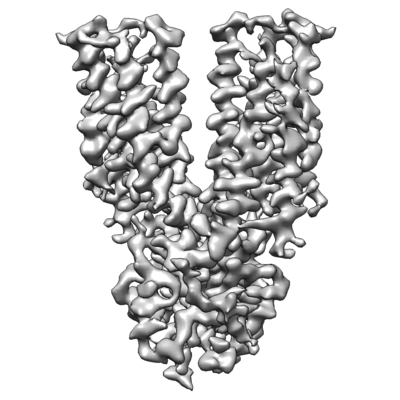 emd_22285.png emd_22285.png | 109.5 KB | ||
| Filedesc metadata | emd-22285.cif.gz | 5.3 KB | ||
| Archive directory |  http://ftp.pdbj.org/pub/emdb/structures/EMD-22285ftp://ftp.pdbj.org/pub/emdb/structures/EMD-22285 http://ftp.pdbj.org/pub/emdb/structures/EMD-22285ftp://ftp.pdbj.org/pub/emdb/structures/EMD-22285 | HTTPS FTP |
-Related structure data
| Related structure data |  6xpdMC  6xpeC  6xpfC C: citing same article ( M: atomic model generated by this map |
|---|---|
| Similar structure data |


-Links
| EMDB pages | EMDB (EBI/PDBe) / EMDataResource |
|---|---|
| Related items in Molecule of the Month |


-Map
| File | Download / File: emd_22285.map.gz / Format: CCP4 / Size: 22.2 MB / Type: IMAGE STORED AS FLOATING POINT NUMBER (4 BYTES) | ||||||||||||||||||||||||||||||||||||||||||||||||||||||||||||||||||||
|---|---|---|---|---|---|---|---|---|---|---|---|---|---|---|---|---|---|---|---|---|---|---|---|---|---|---|---|---|---|---|---|---|---|---|---|---|---|---|---|---|---|---|---|---|---|---|---|---|---|---|---|---|---|---|---|---|---|---|---|---|---|---|---|---|---|---|---|---|---|
| Annotation | Cryo-EM structure of human ZnT8 with two mutations introduced (D110N and D224N), in the outward-facing conformation | ||||||||||||||||||||||||||||||||||||||||||||||||||||||||||||||||||||
| Projections & slices | Image control
Images are generated by Spider. | ||||||||||||||||||||||||||||||||||||||||||||||||||||||||||||||||||||
| Voxel size | X=Y=Z: 0.83 Å | ||||||||||||||||||||||||||||||||||||||||||||||||||||||||||||||||||||
| Density |
| ||||||||||||||||||||||||||||||||||||||||||||||||||||||||||||||||||||
| Symmetry | Space group: 1 | ||||||||||||||||||||||||||||||||||||||||||||||||||||||||||||||||||||
| Details | EMDB XML:
CCP4 map header:
| ||||||||||||||||||||||||||||||||||||||||||||||||||||||||||||||||||||
 Z (Sec.)
 Y (Row.)
 X (Col.)


-Supplemental data
- Sample components
-Entire : Human Znt8 double mutant - D110N and D224N
| Entire | Name: Human Znt8 double mutant - D110N and D224N |
|---|---|
| Components |
|
-Supramolecule #1: Human Znt8 double mutant - D110N and D224N
| Supramolecule | Name: Human Znt8 double mutant - D110N and D224N / type: complex / ID: 1 / Parent: 0 / Macromolecule list: #1 |
|---|---|
| Source (natural) | Organism: Homo sapiens (human) / Location in cell: Insulin secretory granule |
| Molecular weight | Theoretical: 35 KDa |
-Macromolecule #1: Zinc transporter 8
| Macromolecule | Name: Zinc transporter 8 / type: protein_or_peptide / ID: 1 / Number of copies: 2 / Enantiomer: LEVO |
|---|---|
| Source (natural) | Organism: Homo sapiens (human) |
| Molecular weight | Theoretical: 35.08616 KDa |
| Recombinant expression | Organism: Homo sapiens (human) |
| Sequence | String: MYHCHSGSKP TEKGANEYAY AKWKLCSASA ICFIFMIAEV VGGHIAGSLA VVTDAAHLLI NLTSFLLSLF SLWLSSKPPS KRLTFGWHR AEILGALLSI LCIWVVTGVL VYLACERLLY PDYQIQATVM IIVSSCAVAA NIVLTVVLHQ RCLGHNHKEV Q ANASVRAA ...String: MYHCHSGSKP TEKGANEYAY AKWKLCSASA ICFIFMIAEV VGGHIAGSLA VVTDAAHLLI NLTSFLLSLF SLWLSSKPPS KRLTFGWHR AEILGALLSI LCIWVVTGVL VYLACERLLY PDYQIQATVM IIVSSCAVAA NIVLTVVLHQ RCLGHNHKEV Q ANASVRAA FVHALGNLFQ SISVLISALI IYFKPEYKIA DPICTFIFSI LVLASTITIL KDFSILLMEG VPKSLNYSGV KE LILAVDG VLSVHSLHIW SLTMNQVILS AHVATAASRD SQVVRREIAK ALSKSFTMHS LTIQMESPVD QDPDCLFCED PCD UniProtKB: Proton-coupled zinc antiporter SLC30A8 |
-Macromolecule #2: ZINC ION
| Macromolecule | Name: ZINC ION / type: ligand / ID: 2 / Number of copies: 4 / Formula: ZN |
|---|---|
| Molecular weight | Theoretical: 65.409 Da |
-Experimental details
-Structure determination
| Method | cryo EM |
|---|---|
 Processing Processing | single particle reconstruction |
| Aggregation state | particle |
-Sample preparation
| Concentration | 4 mg/mL |
|---|---|
| Buffer | pH: 7.4 |
| Grid | Model: Quantifoil R1.2/1.3 / Material: GOLD / Mesh: 300 / Pretreatment - Type: GLOW DISCHARGE |
| Vitrification | Cryogen name: ETHANE / Chamber humidity: 100 % / Chamber temperature: 277 K / Instrument: FEI VITROBOT MARK IV |
- Electron microscopy
| Microscope | FEI TITAN KRIOS |
|---|---|
| Specialist optics | Phase plate: VOLTA PHASE PLATE / Energy filter - Name: GIF Bioquantum / Energy filter - Slit width: 20 eV |
| Image recording | Film or detector model: GATAN K3 BIOQUANTUM (6k x 4k) / Average electron dose: 60.0 e/Å2 |
| Electron beam | Acceleration voltage: 300 kV / Electron source:  FIELD EMISSION GUN FIELD EMISSION GUN |
| Electron optics | C2 aperture diameter: 70.0 µm / Illumination mode: FLOOD BEAM / Imaging mode: BRIGHT FIELD |
| Sample stage | Specimen holder model: FEI TITAN KRIOS AUTOGRID HOLDER / Cooling holder cryogen: NITROGEN |
| Experimental equipment |  Model: Titan Krios / Image courtesy: FEI Company |
