positive regulation of retinoic acid receptor signaling pathway / Signaling by TCF7L2 mutants / Repression of WNT target genes / oxidoreductase activity, acting on the CH-OH group of donors, NAD or NADP as acceptor / Sensory processing of sound by inner hair cells of the cochlea / white fat cell differentiation / transcription repressor complex / viral genome replication / transcription corepressor binding / transcription coregulator binding ...positive regulation of retinoic acid receptor signaling pathway / Signaling by TCF7L2 mutants / Repression of WNT target genes / oxidoreductase activity, acting on the CH-OH group of donors, NAD or NADP as acceptor / Sensory processing of sound by inner hair cells of the cochlea / white fat cell differentiation / transcription repressor complex / viral genome replication / transcription corepressor binding / transcription coregulator binding / Negative Regulation of CDH1 Gene Transcription / NAD binding / transcription corepressor activity / DNA-binding transcription factor binding / transcription coactivator activity / negative regulation of cell population proliferation / negative regulation of DNA-templated transcription / regulation of transcription by RNA polymerase II / synapse / protein kinase binding / protein-containing complex binding / negative regulation of transcription by RNA polymerase II / positive regulation of transcription by RNA polymerase II / nucleoplasm / identical protein binding / nucleus Similarity search - Function
C-terminal binding protein / : / D-isomer specific 2-hydroxyacid dehydrogenases signature 3. / D-isomer specific 2-hydroxyacid dehydrogenase, NAD-binding domain conserved site / D-isomer specific 2-hydroxyacid dehydrogenase, catalytic domain / D-isomer specific 2-hydroxyacid dehydrogenase, catalytic domain / D-isomer specific 2-hydroxyacid dehydrogenase, NAD-binding domain / D-isomer specific 2-hydroxyacid dehydrogenase, NAD binding domain / NAD(P)-binding domain superfamily Similarity search - Domain/homology
National Institutes of Health/National Institute of General Medical Sciences (NIH/NIGMS)
R01 GM119014
United States
Citation
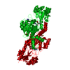
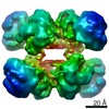
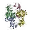
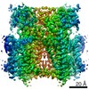
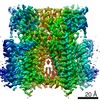
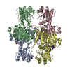
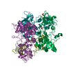
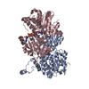
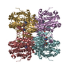
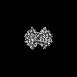
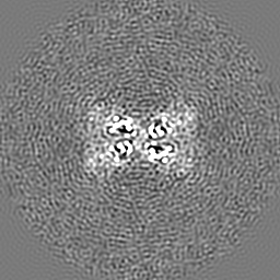
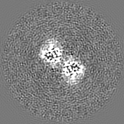
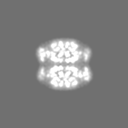
Journal: Structure / Year: 2021 Title: Cryo-EM structure of CtBP2 confirms tetrameric architecture. Authors: Anne M Jecrois / M Michael Dcona / Xiaoyan Deng / Dipankar Bandyopadhyay / Steven R Grossman / Celia A Schiffer / William E Royer / Abstract: C-terminal binding proteins 1 and 2 (CtBP1 and CtBP2) are transcriptional regulators that activate or repress many genes involved in cellular development, apoptosis, and metastasis. NADH-dependent ...C-terminal binding proteins 1 and 2 (CtBP1 and CtBP2) are transcriptional regulators that activate or repress many genes involved in cellular development, apoptosis, and metastasis. NADH-dependent CtBP activation has been implicated in multiple types of cancer and poor patient prognosis. Central to understanding activation of CtBP in oncogenesis is uncovering how NADH triggers protein assembly, what level of assembly occurs, and if oncogenic activity depends upon such assembly. Here, we present the cryoelectron microscopic structures of two different constructs of CtBP2 corroborating that the native state of CtBP2 in the presence of NADH is tetrameric. The physiological relevance of the observed tetramer was demonstrated in cell culture, showing that CtBP tetramer-destabilizing mutants are defective for cell migration, transcriptional repression of E-cadherin, and activation of TIAM1. Together with our cryoelectron microscopy studies, these results highlight the tetramer as the functional oligomeric form of CtBP2.
History
Deposition
Apr 17, 2020
-
Header (metadata) release
Dec 2, 2020
-
Map release
Dec 2, 2020
-
Update
Jun 4, 2025
-
Current status
Jun 4, 2025
Processing site: RCSB / Status: Released
-
Structure visualization
Movie
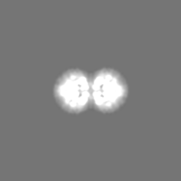
Surface view with section colored by density value
Model: C-flat-1.2/1.3 / Material: COPPER / Mesh: 400 / Pretreatment - Type: GLOW DISCHARGE / Pretreatment - Time: 60 sec. Details: Grid was washed in Ethyl acetate prior to glow-discharge.
Vitrification
Cryogen name: ETHANE / Chamber humidity: 95 % / Chamber temperature: 278.15 K / Instrument: FEI VITROBOT MARK IV / Details: Blotting time: 4s Blotting force: 8 Wait time: 0.
-
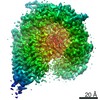
Electron microscopy
Microscope
FEI TALOS ARCTICA
Image recording
Film or detector model: GATAN K3 (6k x 4k) / Number grids imaged: 1 / Number real images: 3405 / Average exposure time: 1.7 sec. / Average electron dose: 37.0 e/Å2
Electron beam
Acceleration voltage: 200 kV / Electron source: FIELD EMISSION GUN
In the structure databanks used in Yorodumi, some data are registered as the other names, "COVID-19 virus" and "2019-nCoV". Here are the details of the virus and the list of structure data.
Jan 31, 2019. EMDB accession codes are about to change! (news from PDBe EMDB page)
EMDB accession codes are about to change! (news from PDBe EMDB page)
The allocation of 4 digits for EMDB accession codes will soon come to an end. Whilst these codes will remain in use, new EMDB accession codes will include an additional digit and will expand incrementally as the available range of codes is exhausted. The current 4-digit format prefixed with “EMD-” (i.e. EMD-XXXX) will advance to a 5-digit format (i.e. EMD-XXXXX), and so on. It is currently estimated that the 4-digit codes will be depleted around Spring 2019, at which point the 5-digit format will come into force.
The EM Navigator/Yorodumi systems omit the EMD- prefix.
Related info.:Q: What is EMD? / ID/Accession-code notation in Yorodumi/EM Navigator
Yorodumi is a browser for structure data from EMDB, PDB, SASBDB, etc.
This page is also the successor to EM Navigator detail page, and also detail information page/front-end page for Omokage search.
The word "yorodu" (or yorozu) is an old Japanese word meaning "ten thousand". "mi" (miru) is to see.
Related info.:EMDB / PDB / SASBDB / Comparison of 3 databanks / Yorodumi Search / Aug 31, 2016. New EM Navigator & Yorodumi / Yorodumi Papers / Jmol/JSmol / Function and homology information / Changes in new EM Navigator and Yorodumi
 Movie
Movie Controller

 Yorodumi
 Open data
 Basic information
 Map data
 Sample
 Keywords
 Function and homology information
 Homo sapiens (human)
 Authors
 United States, 1 items
  Citation
 Structure visualization
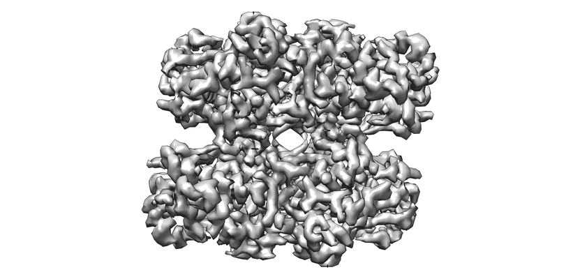
 Downloads & links
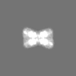
 emd_21811.png
 http://ftp.pdbj.org/pub/emdb/structures/EMD-21811
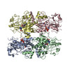

 Z (Sec.)
 Y (Row.)
 X (Col.)


 Sample components

 Processing
 Electron microscopy
 FIELD EMISSION GUN