 Movie
Movie Controller

[English] 日本語
 Yorodumi
- EMDB-21690: Cryo-EM structure of apo-Photosystem II from Synechocystis sp. PC... -
+ Open data

- Basic information
| Entry | Database: EMDB / ID: EMD-21690 | |||||||||
|---|---|---|---|---|---|---|---|---|---|---|
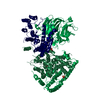
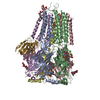
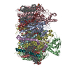
| Title | Cryo-EM structure of apo-Photosystem II from Synechocystis sp. PCC 6803 | |||||||||
 Map data Map data | apo-photosystem II | |||||||||
 Sample Sample |
| |||||||||
 Keywords Keywords | Photosystem / oxidoreductase / water-splitting / photoactivation / PHOTOSYNTHESIS | |||||||||
| Function / homology |  Function and homology information Function and homology informationplasma membrane-derived thylakoid photosystem II / thylakoid / oxygen evolving activity / photosystem II reaction center / photosystem II / photosynthetic electron transport chain / oxidoreductase activity, acting on diphenols and related substances as donors, oxygen as acceptor / response to herbicide / photosystem II / plasma membrane-derived thylakoid membrane ...plasma membrane-derived thylakoid photosystem II / thylakoid / oxygen evolving activity / photosystem II reaction center / photosystem II / photosynthetic electron transport chain / oxidoreductase activity, acting on diphenols and related substances as donors, oxygen as acceptor / response to herbicide / photosystem II / plasma membrane-derived thylakoid membrane / photosynthetic electron transport in photosystem II / chlorophyll binding / photosynthesis, light reaction / phosphate ion binding / photosynthesis / electron transfer activity / protein stabilization / iron ion binding / heme binding / metal ion binding / identical protein binding Similarity search - Function | |||||||||
| Biological species |  | |||||||||
| Method | single particle reconstruction / cryo EM / Resolution: 2.58 Å | |||||||||
 Authors Authors | Gisriel CJ | |||||||||
| Funding support |  United States, 1 items United States, 1 items
| |||||||||
 Citation Citation | Journal: Joule / Year: 2020 Title: Cryo-EM Structure of Monomeric Photosystem II from Synechocystis sp. PCC 6803 Lacking the Water-Oxidation Complex Authors: Gisriel CJ / Zhou K / Huang H / Debus RJ / Xiong Y / Brudvig GW | |||||||||
| History |
|
- Structure visualization
| Movie |
Movie viewer |
|---|---|
| Structure viewer | EM map: SurfViewMolmilJmol/JSmol |
| Supplemental images |

- Downloads & links
-EMDB archive
| Map data | emd_21690.map.gz | 59.9 MB | EMDB map data format | |
|---|---|---|---|---|
| Header (meta data) | emd-21690-v30.xmlemd-21690.xml | 28.8 KB 28.8 KB | Display Display | EMDB header |
| FSC (resolution estimation) | emd_21690_fsc.xml | 9.1 KB | Display | FSC data file |
| Images |  emd_21690.png emd_21690.png | 22.4 KB | ||
| Filedesc metadata | emd-21690.cif.gz | 7.8 KB | ||
| Archive directory |  http://ftp.pdbj.org/pub/emdb/structures/EMD-21690ftp://ftp.pdbj.org/pub/emdb/structures/EMD-21690 http://ftp.pdbj.org/pub/emdb/structures/EMD-21690ftp://ftp.pdbj.org/pub/emdb/structures/EMD-21690 | HTTPS FTP |
-Related structure data
| Related structure data |  6wj6MC M: atomic model generated by this map C: citing same article ( |
|---|---|
| Similar structure data |


-Links
| EMDB pages | EMDB (EBI/PDBe) / EMDataResource |
|---|---|
| Related items in Molecule of the Month |


-Map
| File | Download / File: emd_21690.map.gz / Format: CCP4 / Size: 64 MB / Type: IMAGE STORED AS FLOATING POINT NUMBER (4 BYTES) | ||||||||||||||||||||||||||||||||||||||||||||||||||||||||||||
|---|---|---|---|---|---|---|---|---|---|---|---|---|---|---|---|---|---|---|---|---|---|---|---|---|---|---|---|---|---|---|---|---|---|---|---|---|---|---|---|---|---|---|---|---|---|---|---|---|---|---|---|---|---|---|---|---|---|---|---|---|---|
| Annotation | apo-photosystem II | ||||||||||||||||||||||||||||||||||||||||||||||||||||||||||||
| Projections & slices | Image control
Images are generated by Spider. | ||||||||||||||||||||||||||||||||||||||||||||||||||||||||||||
| Voxel size | X=Y=Z: 1.18125 Å | ||||||||||||||||||||||||||||||||||||||||||||||||||||||||||||
| Density |
| ||||||||||||||||||||||||||||||||||||||||||||||||||||||||||||
| Symmetry | Space group: 1 | ||||||||||||||||||||||||||||||||||||||||||||||||||||||||||||
| Details | EMDB XML:
CCP4 map header:
| ||||||||||||||||||||||||||||||||||||||||||||||||||||||||||||
 Z (Sec.)
 Y (Row.)
 X (Col.)


-Supplemental data
- Sample components
+Entire : apo-Photosystem II
+Supramolecule #1: apo-Photosystem II
+Macromolecule #1: Photosystem II protein D1 2
+Macromolecule #2: Photosystem II CP47 reaction center protein
+Macromolecule #3: Photosystem II CP43 reaction center protein
+Macromolecule #4: Photosystem II D2 protein
+Macromolecule #5: Cytochrome b559 subunit alpha
+Macromolecule #6: Cytochrome b559 subunit beta
+Macromolecule #7: Photosystem II reaction center protein H
+Macromolecule #8: Photosystem II reaction center protein I
+Macromolecule #9: Photosystem II reaction center protein K
+Macromolecule #10: Photosystem II reaction center protein L
+Macromolecule #11: Photosystem II reaction center protein M
+Macromolecule #12: Photosystem II reaction center protein T
+Macromolecule #13: Photosystem II reaction center X protein
+Macromolecule #14: FE (II) ION
+Macromolecule #15: CHLOROPHYLL A

+Macromolecule #16: PHEOPHYTIN A

+Macromolecule #17: BETA-CAROTENE

+Macromolecule #18: 1,2-DI-O-ACYL-3-O-[6-DEOXY-6-SULFO-ALPHA-D-GLUCOPYRANOSYL]-SN-GLYCEROL

+Macromolecule #19: 2,3-DIMETHYL-5-(3,7,11,15,19,23,27,31,35-NONAMETHYL-2,6,10,14,18,...
+Macromolecule #20: 1,2-DISTEAROYL-MONOGALACTOSYL-DIGLYCERIDE

+Macromolecule #21: DODECYL-BETA-D-MALTOSIDE

+Macromolecule #22: DIGALACTOSYL DIACYL GLYCEROL (DGDG)

+Macromolecule #23: CHLORIDE ION
+Macromolecule #24: BICARBONATE ION

+Macromolecule #25: 1,2-DIPALMITOYL-PHOSPHATIDYL-GLYCEROLE

+Macromolecule #26: PROTOPORPHYRIN IX CONTAINING FE

+Macromolecule #27: N-FORMYLMETHIONINE
+Macromolecule #28: water

-Experimental details
-Structure determination
| Method | cryo EM |
|---|---|
 Processing Processing | single particle reconstruction |
| Aggregation state | particle |
-Sample preparation
| Concentration | 0.025 mg/mL |
|---|---|
| Buffer | pH: 7 Details: 10 mM HEPES pH 7.0 100 mM NaCl 0.03% beta-dodecylmaltoside |
| Grid | Model: C-flat-2/1 / Material: COPPER / Mesh: 300 / Support film - Material: CARBON / Support film - topology: HOLEY / Pretreatment - Type: GLOW DISCHARGE / Pretreatment - Time: 30 sec. |
| Vitrification | Cryogen name: ETHANE / Chamber humidity: 100 % / Chamber temperature: 298 K / Instrument: FEI VITROBOT MARK IV |
| Details | Monodisperse single particles |
- Electron microscopy
| Microscope | FEI TITAN KRIOS |
|---|---|
| Image recording | Film or detector model: GATAN K2 SUMMIT (4k x 4k) / Average electron dose: 48.07 e/Å2 |
| Electron beam | Acceleration voltage: 300 kV / Electron source:  FIELD EMISSION GUN FIELD EMISSION GUN |
| Electron optics | Illumination mode: FLOOD BEAM / Imaging mode: BRIGHT FIELD |
| Experimental equipment |  Model: Titan Krios / Image courtesy: FEI Company |

