 Movie
Movie Controller

[English] 日本語
 Yorodumi
- EMDB-21611: Asymmetric vertex of the clathrin minicoat cage, subparticle refi... -
+ Open data

- Basic information
| Entry | Database: EMDB / ID: EMD-21611 | |||||||||
|---|---|---|---|---|---|---|---|---|---|---|
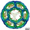
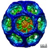
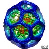
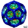
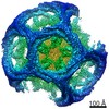
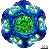
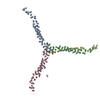
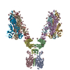
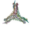
| Title | Asymmetric vertex of the clathrin minicoat cage, subparticle refinement of the clathrin layer | |||||||||
 Map data Map data | Sharpened asymmetric vertex | |||||||||
 Sample Sample |
| |||||||||
 Keywords Keywords | Clathrin coated vesicle / clathrin heavy chain / clathrin light chain / clathrin cage / ENDOCYTOSIS | |||||||||
| Function / homology |  Function and homology information Function and homology informationRetrograde neurotrophin signalling / Recycling pathway of L1 / WNT5A-dependent internalization of FZD4 / WNT5A-dependent internalization of FZD2, FZD5 and ROR2 / LDL clearance / Gap junction degradation / Formation of annular gap junctions / clathrin vesicle coat / RHOU GTPase cycle / RHOV GTPase cycle ...Retrograde neurotrophin signalling / Recycling pathway of L1 / WNT5A-dependent internalization of FZD4 / WNT5A-dependent internalization of FZD2, FZD5 and ROR2 / LDL clearance / Gap junction degradation / Formation of annular gap junctions / clathrin vesicle coat / RHOU GTPase cycle / RHOV GTPase cycle / clathrin coat of trans-Golgi network vesicle / Golgi Associated Vesicle Biogenesis / clathrin light chain binding / Lysosome Vesicle Biogenesis / negative regulation of hyaluronan biosynthetic process / clathrin complex / MHC class II antigen presentation / VLDLR internalisation and degradation / clathrin coat / clathrin coat of coated pit / clathrin coat disassembly / Cargo recognition for clathrin-mediated endocytosis / clathrin heavy chain binding / clathrin-coated endocytic vesicle / membrane coat / clathrin coat assembly / Clathrin-mediated endocytosis / clathrin-dependent endocytosis / arrestin family protein binding / receptor-mediated endocytosis / intracellular protein transport / autophagy / spindle / disordered domain specific binding / melanosome / mitotic cell cycle / synaptic vesicle membrane / protein domain specific binding / cell division / structural molecule activity / mitochondrion / identical protein binding / plasma membrane Similarity search - Function | |||||||||
| Biological species |  | |||||||||
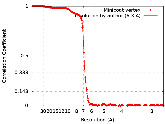
| Method | single particle reconstruction / cryo EM / Resolution: 6.3 Å | |||||||||
 Authors Authors | Paraan M / Mendez J / Sharum S / Kurtin D / He H / Stagg S | |||||||||
| Funding support |  United States, 1 items United States, 1 items
| |||||||||
 Citation Citation | Journal: Sci Adv / Year: 2020 Title: The structures of natively assembled clathrin-coated vesicles. Authors: Mohammadreza Paraan / Joshua Mendez / Savanna Sharum / Danielle Kurtin / Huan He / Scott M Stagg / Abstract: Clathrin-coated vesicles mediate trafficking of proteins and nutrients in the cell and between organelles. Proteins included in the clathrin-coated vesicles (CCVs) category include clathrin heavy ...Clathrin-coated vesicles mediate trafficking of proteins and nutrients in the cell and between organelles. Proteins included in the clathrin-coated vesicles (CCVs) category include clathrin heavy chain (CHC), clathrin light chain (CLC), and a variety of adaptor protein complexes. Much is known about the structures of the individual CCV components, but data are lacking about the structures of the fully assembled complexes together with membrane and in complex with cargo. Here, we determined the structures of natively assembled CCVs in a variety of geometries. We show that the adaptor β2 appendages crosslink adjacent CHC β-propellers and that the appendage densities are enriched in CCV hexagonal faces. We resolve how adaptor protein 2 and other associated factors in hexagonal faces form an assembly hub with an extensive web of interactions between neighboring β-propellers and propose a structural model that explains how adaptor binding can direct the formation of pentagonal and hexagonal faces. | |||||||||
| History |
|
- Structure visualization
| Movie |
Movie viewer |
|---|---|
| Structure viewer | EM map: SurfViewMolmilJmol/JSmol |
| Supplemental images |

- Downloads & links
-EMDB archive
| Map data | emd_21611.map.gz | 44.6 MB | EMDB map data format | |
|---|---|---|---|---|
| Header (meta data) | emd-21611-v30.xmlemd-21611.xml | 32.7 KB 32.7 KB | Display Display | EMDB header |
| FSC (resolution estimation) | emd_21611_fsc.xml | 21 KB | Display | FSC data file |
| Images |  emd_21611.png emd_21611.png | 115.8 KB | ||
| Masks | emd_21611_msk_1.map | 824 MB | Mask map | |
| Filedesc metadata | emd-21611.cif.gz | 7.6 KB | ||
| Others | emd_21611_additional.map.gzemd_21611_half_map_1.map.gzemd_21611_half_map_2.map.gz | 764 MB 523.2 MB 523.2 MB | ||
| Archive directory |  http://ftp.pdbj.org/pub/emdb/structures/EMD-21611ftp://ftp.pdbj.org/pub/emdb/structures/EMD-21611 http://ftp.pdbj.org/pub/emdb/structures/EMD-21611ftp://ftp.pdbj.org/pub/emdb/structures/EMD-21611 | HTTPS FTP |
-Related structure data
| Related structure data |  6wcjMC C: citing same article ( M: atomic model generated by this map |
|---|---|
| Similar structure data |


-Links
| EMDB pages | EMDB (EBI/PDBe) / EMDataResource |
|---|---|
| Related items in Molecule of the Month |

-Map
| File | Download / File: emd_21611.map.gz / Format: CCP4 / Size: 824 MB / Type: IMAGE STORED AS FLOATING POINT NUMBER (4 BYTES) | ||||||||||||||||||||||||||||||||||||||||||||||||||||||||||||||||||||
|---|---|---|---|---|---|---|---|---|---|---|---|---|---|---|---|---|---|---|---|---|---|---|---|---|---|---|---|---|---|---|---|---|---|---|---|---|---|---|---|---|---|---|---|---|---|---|---|---|---|---|---|---|---|---|---|---|---|---|---|---|---|---|---|---|---|---|---|---|---|
| Annotation | Sharpened asymmetric vertex | ||||||||||||||||||||||||||||||||||||||||||||||||||||||||||||||||||||
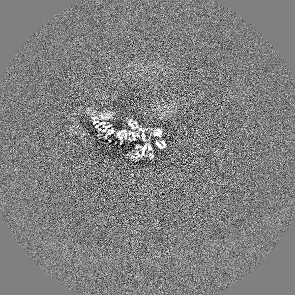
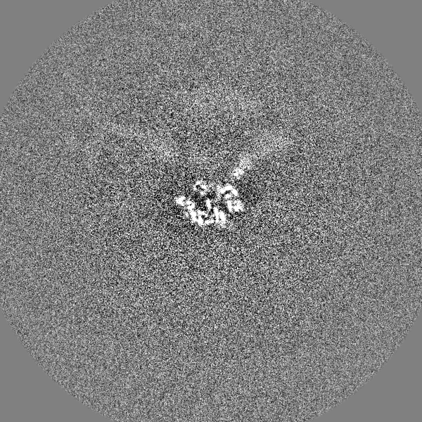
| Projections & slices | Image control
Images are generated by Spider. | ||||||||||||||||||||||||||||||||||||||||||||||||||||||||||||||||||||
| Voxel size | X=Y=Z: 1.323 Å | ||||||||||||||||||||||||||||||||||||||||||||||||||||||||||||||||||||
| Density |
| ||||||||||||||||||||||||||||||||||||||||||||||||||||||||||||||||||||
| Symmetry | Space group: 1 | ||||||||||||||||||||||||||||||||||||||||||||||||||||||||||||||||||||
| Details | EMDB XML:
CCP4 map header:
| ||||||||||||||||||||||||||||||||||||||||||||||||||||||||||||||||||||
 Z (Sec.)
 Y (Row.)
 X (Col.)


-Supplemental data
-Mask #1
| File | emd_21611_msk_1.map | ||||||||||||
|---|---|---|---|---|---|---|---|---|---|---|---|---|---|
| Projections & Slices |
| ||||||||||||
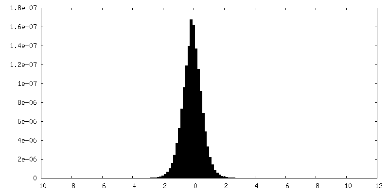
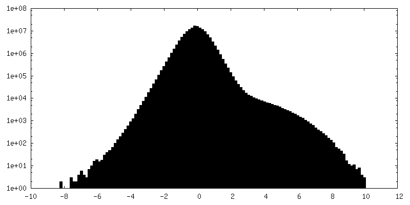
| Density Histograms |


-Additional map: Nonsharpened asymmetric vertex
| File | emd_21611_additional.map | ||||||||||||
|---|---|---|---|---|---|---|---|---|---|---|---|---|---|
| Annotation | Nonsharpened asymmetric vertex | ||||||||||||
| Projections & Slices |
| ||||||||||||
| Density Histograms |


-Half map: Unmasked half map
| File | emd_21611_half_map_1.map | ||||||||||||
|---|---|---|---|---|---|---|---|---|---|---|---|---|---|
| Annotation | Unmasked half map | ||||||||||||
| Projections & Slices |
| ||||||||||||
| Density Histograms |


-Half map: Unmasked half map
| File | emd_21611_half_map_2.map | ||||||||||||
|---|---|---|---|---|---|---|---|---|---|---|---|---|---|
| Annotation | Unmasked half map | ||||||||||||
| Projections & Slices |
| ||||||||||||
| Density Histograms |


- Sample components
-Entire : Natively assembled clathrin coated vesicles
| Entire | Name: Natively assembled clathrin coated vesicles |
|---|---|
| Components |
|
-Supramolecule #1: Natively assembled clathrin coated vesicles
| Supramolecule | Name: Natively assembled clathrin coated vesicles / type: complex / ID: 1 / Parent: 0 / Macromolecule list: all |
|---|---|
| Source (natural) | Organism: |
-Macromolecule #1: Clathrin heavy chain 1
| Macromolecule | Name: Clathrin heavy chain 1 / type: protein_or_peptide / ID: 1 / Number of copies: 9 / Enantiomer: LEVO |
|---|---|
| Source (natural) | Organism: |
| Molecular weight | Theoretical: 191.800312 KDa |
| Sequence | String: MAQILPIRFQ EHLQLQNLGI NPANIGFSTL TMESDKFICI REKVGEQAQV VIIDMNDPSN PIRRPISADS AIMNPASKVI ALKAGKTLQ IFNIEMKSKM KAHTMTDDVT FWKWISLNTV ALVTDNAVYH WSMEGESQPV KMFDRHSSLA GCQIINYRTD A KQKWLLLT ...String: MAQILPIRFQ EHLQLQNLGI NPANIGFSTL TMESDKFICI REKVGEQAQV VIIDMNDPSN PIRRPISADS AIMNPASKVI ALKAGKTLQ IFNIEMKSKM KAHTMTDDVT FWKWISLNTV ALVTDNAVYH WSMEGESQPV KMFDRHSSLA GCQIINYRTD A KQKWLLLT GISAQQNRVV GAMQLYSVDR KVSQPIEGHA ASFAQFKMEG NAEESTLFCF AVRGQAGGKL HIIEVGTPPT GN QPFPKKA VDVFFPPEAQ NDFPVAMQIS EKHDVVFLIT KYGYIHLYDL ETGTCIYMNR ISGETIFVTA PHEATAGIIG VNR KGQVLS VCVEEENIIP YITNVLQNPD LALRMAVRNN LAGAEELFAR KFNALFAQGN YSEAAKVAAN APKGILRTPD TIRR FQSVP AQPGQTSPLL QYFGILLDQG QLNKYESLEL CRPVLQQGRK QLLEKWLKED KLECSEELGD LVKSVDPTLA LSVYL RANV PNKVIQCFAE TGQVQKIVLY AKKVGYTPDW IFLLRNVMRI SPDQGQQFAQ MLVQDEEPLA DITQIVDVFM EYNLIQ QCT AFLLDALKNN RPSEGPLQTR LLEMNLMHAP QVADAILGNQ MFTHYDRAHI AQLCEKAGLL QRALEHFTDL YDIKRAV VH THLLNPEWLV NYFGSLSVED SLECLRAMLS ANIRQNLQIC VQVASKYHEQ LSTQSLIELF ESFKSFEGLF YFLGSIVN F SQDPDVHFKY IQAACKTGQI KEVERICRES NCYDPERVKN FLKEAKLTDQ LPLIIVCDRF DFVHDLVLYL YRNNLQKYI EIYVQKVNPS RLPVVIGGLL DVDCSEDVIK NLILVVRGQF STDELVAEVE KRNRLKLLLP WLEARIHEGC EEPATHNALA KIYIDSNNN PERFLRENPY YDSRVVGKYC EKRDPHLACV AYERGQCDLE LINVCNENSL FKSLSRYLVR RKDPELWGSV L LESNPYRR PLIDQVVQTA LSETQDPEEV SVTVKAFMTA DLPNELIELL EKIVLDNSVF SEHRNLQNLL ILTAIKADRT RV MEYINRL DNYDAPDIAN IAISNELFEE AFAIFRKFDV NTSAVQVLIE HIGNLDRAYE FAERCNEPAV WSQLAKAQLQ KGM VKEAID SYIKADDPSS YMEVVQAANT SGNWEELVKY LQMARKKARE SYVETELIFA LAKTNRLAEL EEFINGPNNA HIQQ VGDRC YDEKMYDAAK LLYNNVSNFG RLASTLVHLG EYQAAVDGAR KANSTRTWKE VCFACVDGKE FRLAQMCGLH IVVHA DELE ELINYYQDRG YFEELITMLE AALGLERAHM GMFTELAILY SKFKPQKMRE HLELFWSRVN IPKVLRAAEQ AHLWAE LVF LYDKYEEYDN AIITMMNHPT DAWKEGQFKD IITKVANVEL YYRAIQFYLE FKPLLLNDLL MVLSPRLDHT RAVNYFS KV KQLPLVKPYL RSVQNHNNKS VNESLNNLFI TEEDYQALRT SIDAYDNFDN ISLAQRLEKH ELIEFRRIAA YLFKGNNR W KQSVELCKKD SLYKDAMQYA SESKDTELAE ELLQWFLQEE KRECFGACLF TCYDLLRPDV VLETAWRHNI MDFAMPYFI QVMKEYLTKV DKLDASESLR KEEEQATETQ PIVYGQPQLM LTAGPSVAVP PQAPFGYGYT APAYGQPQPG FGYSM UniProtKB: Clathrin heavy chain 1 |
-Macromolecule #2: Clathrin light chain B
| Macromolecule | Name: Clathrin light chain B / type: protein_or_peptide / ID: 2 / Number of copies: 6 / Enantiomer: LEVO |
|---|---|
| Source (natural) | Organism: |
| Molecular weight | Theoretical: 25.109396 KDa |
| Sequence | String: MADDFGFFSS SESGAPEAAE EDPAAAFLAQ QESEIAGIEN DEGFGAPAGS QGGLAQPGPA SGASEDMGAT VNGDVFQEAN GPADGYAAI AQADRLTQEP ESIRKWREEQ RKRLQELDAA SKVMEQEWRE KAKKDLEEWN QRQSEQVEKN KINNRIADKA F YQQPDADI ...String: MADDFGFFSS SESGAPEAAE EDPAAAFLAQ QESEIAGIEN DEGFGAPAGS QGGLAQPGPA SGASEDMGAT VNGDVFQEAN GPADGYAAI AQADRLTQEP ESIRKWREEQ RKRLQELDAA SKVMEQEWRE KAKKDLEEWN QRQSEQVEKN KINNRIADKA F YQQPDADI IGYVASEEAF VKESKEETPG TEWEKVAQLC DFNPKSSKQC KDVSRLRSVL MSLKQTPLSR UniProtKB: Clathrin light chain B |
-Experimental details
-Structure determination
| Method | cryo EM |
|---|---|
 Processing Processing | single particle reconstruction |
| Aggregation state | particle |
-Sample preparation
| Concentration | 6 mg/mL | ||||||||||||
|---|---|---|---|---|---|---|---|---|---|---|---|---|---|
| Buffer | pH: 6.7 Component:
| ||||||||||||
| Grid | Model: C-flat-2/2 4C | ||||||||||||
| Vitrification | Cryogen name: ETHANE / Chamber humidity: 100 % / Chamber temperature: 281 K / Instrument: FEI VITROBOT MARK IV |
- Electron microscopy
| Microscope | FEI TITAN KRIOS |
|---|---|
| Image recording | Film or detector model: GATAN K3 (6k x 4k) / Number grids imaged: 1 / Number real images: 800 / Average electron dose: 50.0 e/Å2 |
| Electron beam | Acceleration voltage: 300 kV / Electron source:  FIELD EMISSION GUN FIELD EMISSION GUN |
| Electron optics | C2 aperture diameter: 70.0 µm / Illumination mode: FLOOD BEAM / Imaging mode: BRIGHT FIELD / Cs: 2.7 mm / Nominal defocus max: 5.0 µm / Nominal defocus min: 3.0 µm / Nominal magnification: 35000 |
| Sample stage | Specimen holder model: FEI TITAN KRIOS AUTOGRID HOLDER / Cooling holder cryogen: NITROGEN |
| Experimental equipment |  Model: Titan Krios / Image courtesy: FEI Company |
+Image processing
-Atomic model buiding 1
| Initial model |
| ||||||||
|---|---|---|---|---|---|---|---|---|---|
| Refinement | Protocol: AB INITIO MODEL | ||||||||
| Output model | PDB-6wcj: |
-Atomic model buiding 2
| Initial model |
| ||||||||
|---|---|---|---|---|---|---|---|---|---|
| Refinement | Protocol: AB INITIO MODEL | ||||||||
| Output model | PDB-6wcj: |
-Atomic model buiding 3
| Initial model |
| ||||||||||||||||||||||||||||||||
|---|---|---|---|---|---|---|---|---|---|---|---|---|---|---|---|---|---|---|---|---|---|---|---|---|---|---|---|---|---|---|---|---|---|
| Refinement | Protocol: FLEXIBLE FIT | ||||||||||||||||||||||||||||||||
| Output model | PDB-6wcj: |
