National Institutes of Health/National Institute of General Medical Sciences (NIH/NIGMS)
GM108753
United States
Citation
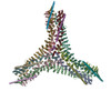
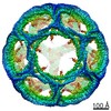
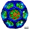
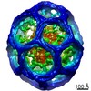
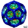
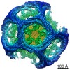
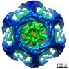
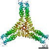
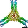
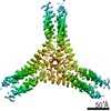
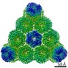
Journal: Sci Adv / Year: 2020 Title: The structures of natively assembled clathrin-coated vesicles. Authors: Mohammadreza Paraan / Joshua Mendez / Savanna Sharum / Danielle Kurtin / Huan He / Scott M Stagg / Abstract: Clathrin-coated vesicles mediate trafficking of proteins and nutrients in the cell and between organelles. Proteins included in the clathrin-coated vesicles (CCVs) category include clathrin heavy ...Clathrin-coated vesicles mediate trafficking of proteins and nutrients in the cell and between organelles. Proteins included in the clathrin-coated vesicles (CCVs) category include clathrin heavy chain (CHC), clathrin light chain (CLC), and a variety of adaptor protein complexes. Much is known about the structures of the individual CCV components, but data are lacking about the structures of the fully assembled complexes together with membrane and in complex with cargo. Here, we determined the structures of natively assembled CCVs in a variety of geometries. We show that the adaptor β2 appendages crosslink adjacent CHC β-propellers and that the appendage densities are enriched in CCV hexagonal faces. We resolve how adaptor protein 2 and other associated factors in hexagonal faces form an assembly hub with an extensive web of interactions between neighboring β-propellers and propose a structural model that explains how adaptor binding can direct the formation of pentagonal and hexagonal faces.
#256 - Apr 2021 SARS-CoV-2 Spike and Antibodies similarity (1)
-
Assembly
Deposited unit
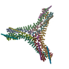
A: Clathrin heavy chain 1 B: Clathrin light chain B C: Clathrin heavy chain 1 E: Clathrin light chain B D: Clathrin heavy chain 1 F: Clathrin light chain B O: Clathrin light chain B L: Clathrin heavy chain 1 M: Clathrin heavy chain 1 J: Clathrin light chain B G: Clathrin heavy chain 1 H: Clathrin heavy chain 1 N: Clathrin light chain B I: Clathrin heavy chain 1 K: Clathrin heavy chain 1
Evidence: mass spectrometry, Using mass spectrometry, the presence of adaptors necessary for the assembly of clathrin coated vesicles in vivo was confirmed.
Type
Name
Symmetry operation
Number
identity operation
1_555
1
Buried area
50200 Å2
ΔGint
-114 kcal/mol
Surface area
264910 Å2
-
Components
#1: Antibody
Clathrinheavychain1
Mass: 191800.312 Da / Num. of mol.: 9 / Source method: isolated from a natural source / Source: (natural) Bos taurus (domestic cattle) / References: UniProt: P49951
#2: Protein
ClathrinlightchainB / Lcb
Mass: 25109.396 Da / Num. of mol.: 6 / Source method: isolated from a natural source / Source: (natural) Bos taurus (domestic cattle) / References: UniProt: P04975
-
Experimental details
-
Experiment
Experiment
Method: ELECTRON MICROSCOPY
EM experiment
Aggregation state: PARTICLE / 3D reconstruction method: single particle reconstruction
Electron dose: 50 e/Å2 / Film or detector model: GATAN K3 (6k x 4k) / Num. of grids imaged: 1 / Num. of real images: 800
-
Processing
Software
Name
Version
Classification
phenix.real_space_refine
1.17_3644
refinement
PHENIX
1.17_3644
refinement
EM software
ID
Name
Version
Category
Fitting-ID
1
RELION
3
particleselection
2
Leginon
imageacquisition
4
cisTEM
1
CTFcorrection
5
CTFFIND
4
CTFcorrection
6
RELION
3
CTFcorrection
9
Rosetta
modelfitting
1
11
cryoSPARC
2
initialEulerassignment
12
cisTEM
1
finalEulerassignment
13
RELION
3
finalEulerassignment
15
cisTEM
1
3Dreconstruction
35
PHENIX
modelrefinement
1
63
Rosetta
modelfitting
2
64
PHENIX
modelrefinement
2
92
Rosetta
modelfitting
3
93
PHENIX
modelrefinement
3
CTF correction
Type: PHASE FLIPPING AND AMPLITUDE CORRECTION
Particle selection
Num. of particles selected: 360000 Details: These are subparticle images that were extracted from particle images. Each minicoat particle has 24 asymmetric vertices. From a total of 15000 minicoat particles, 360000 vertex subparticles ...Details: These are subparticle images that were extracted from particle images. Each minicoat particle has 24 asymmetric vertices. From a total of 15000 minicoat particles, 360000 vertex subparticles were extracted. The position of subparticles was calculated by the localized reconstruction script in scipion, and the subparticle images were extracted with RELION extraction tools.
Symmetry
Point symmetry: C1 (asymmetric)
3D reconstruction
Resolution: 6.3 Å / Resolution method: FSC 0.143 CUT-OFF / Num. of particles: 305413 Details: After the final refinement in cisTEM, half-maps were generated in cisTEM and the FSC was calculated with EMAN FSC tools. Symmetry type: POINT
In the structure databanks used in Yorodumi, some data are registered as the other names, "COVID-19 virus" and "2019-nCoV". Here are the details of the virus and the list of structure data.
Jan 31, 2019. EMDB accession codes are about to change! (news from PDBe EMDB page)
EMDB accession codes are about to change! (news from PDBe EMDB page)
The allocation of 4 digits for EMDB accession codes will soon come to an end. Whilst these codes will remain in use, new EMDB accession codes will include an additional digit and will expand incrementally as the available range of codes is exhausted. The current 4-digit format prefixed with “EMD-” (i.e. EMD-XXXX) will advance to a 5-digit format (i.e. EMD-XXXXX), and so on. It is currently estimated that the 4-digit codes will be depleted around Spring 2019, at which point the 5-digit format will come into force.
The EM Navigator/Yorodumi systems omit the EMD- prefix.
Related info.:Q: What is EMD? / ID/Accession-code notation in Yorodumi/EM Navigator
Yorodumi is a browser for structure data from EMDB, PDB, SASBDB, etc.
This page is also the successor to EM Navigator detail page, and also detail information page/front-end page for Omokage search.
The word "yorodu" (or yorozu) is an old Japanese word meaning "ten thousand". "mi" (miru) is to see.
Related info.:EMDB / PDB / SASBDB / Comparison of 3 databanks / Yorodumi Search / Aug 31, 2016. New EM Navigator & Yorodumi / Yorodumi Papers / Jmol/JSmol / Function and homology information / Changes in new EM Navigator and Yorodumi
 Movie
Movie Controller


 Open data
 Basic information
 Components
 Keywords
 Function and homology information
 Authors
 United States, 1items
  Citation
 Structure visualization
 Downloads & links
 Other downloads


 PDBj
PDBj


 Assembly
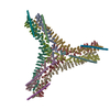
 Sample preparation
 Electron microscopy imaging
 FIELD EMISSION GUN / Accelerating voltage: 300 kV / Illumination mode: FLOOD BEAM
 Processing