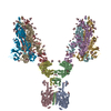
A: Clathrin heavy chain 1 B: Clathrin heavy chain 1 C: Clathrin heavy chain 1 D: Clathrin light chain B E: Clathrin light chain B F: Clathrin light chain B
Mass: 72180.977 Da / Num. of mol.: 3 Source method: isolated from a genetically manipulated source Source: (gene. exp.) Bos taurus (domestic cattle) / Gene: CLTC / Production host: Escherichia coli (E. coli) / Strain (production host): BL21(DE3) / References: UniProt: P49951
#2: Protein
ClathrinlightchainB / Lcb
Mass: 19147.379 Da / Num. of mol.: 3 Source method: isolated from a genetically manipulated source Source: (gene. exp.) Bos taurus (domestic cattle) / Gene: CLTB, CLTLB / Production host: Escherichia coli (E. coli) / Strain (production host): BL21(DE3) / References: UniProt: P04975
Sequence details
FULL-LENGTH CLATHRIN LIGHT CHAIN B WAS USED IN THE CRYSTALLIZATION EXPERIMENT (UNIPROT ID P04975, ...FULL-LENGTH CLATHRIN LIGHT CHAIN B WAS USED IN THE CRYSTALLIZATION EXPERIMENT (UNIPROT ID P04975, 228 RESIDUES).
-
Experimental details
-
Experiment
Experiment
Method: X-RAY DIFFRACTION / Number of used crystals: 1
-
Sample preparation
Crystal grow
Temperature: 298 K / Method: vapor diffusion, hanging drop Details: 200mM citrate, 16-22% glycerol, 2% trifluoroethanol, vapor diffusion, hanging drop, temperature 298K PH range: 4.0-5.0
-
Data collection
Diffraction source
Source: SYNCHROTRON / Site: ALS / Beamline: 8.3.1 / Wavelength: 1.017 Å
Detector
Detector: CCD / Date: Mar 3, 2002
Radiation
Protocol: SINGLE WAVELENGTH / Scattering type: x-ray
Radiation wavelength
Wavelength: 1.017 Å / Relative weight: 1
Reflection
Resolution: 7.9→250 Å / Num. obs: 10700 / Redundancy: 9 % / Rsym value: 0.075 / Net I/σ(I): 20
Method to determine structure: MOLECULAR REPLACEMENT / Resolution: 7.94→100 Å / Occupancy max: 1 / Occupancy min: 1 / σ(F): 0 Details: Due to the low resolution the side chain positions of UNK residues are undetermined though they were present in the phasing and refinement of the structure. Reflection data was processed ...Details: Due to the low resolution the side chain positions of UNK residues are undetermined though they were present in the phasing and refinement of the structure. Reflection data was processed using multiple resolution cutoffs. Initially, the highest resolution was 8.3A. To determine the best resolution for structure determination, maps were calculated with molecular replacement phases and manually inspected for increased structural details. For this manual determination of the resolution data was processed up to 5A resolution in an attempt to gain the highest resolution possible. The reflection data used for structure refinement was cutoff at 7.9A. Reflection data processed to a variety of other high resolution cutoffs can be obtained by sending a request to the authors.
In the structure databanks used in Yorodumi, some data are registered as the other names, "COVID-19 virus" and "2019-nCoV". Here are the details of the virus and the list of structure data.
Jan 31, 2019. EMDB accession codes are about to change! (news from PDBe EMDB page)
EMDB accession codes are about to change! (news from PDBe EMDB page)
The allocation of 4 digits for EMDB accession codes will soon come to an end. Whilst these codes will remain in use, new EMDB accession codes will include an additional digit and will expand incrementally as the available range of codes is exhausted. The current 4-digit format prefixed with “EMD-” (i.e. EMD-XXXX) will advance to a 5-digit format (i.e. EMD-XXXXX), and so on. It is currently estimated that the 4-digit codes will be depleted around Spring 2019, at which point the 5-digit format will come into force.
The EM Navigator/Yorodumi systems omit the EMD- prefix.
Related info.:Q: What is EMD? / ID/Accession-code notation in Yorodumi/EM Navigator
Yorodumi is a browser for structure data from EMDB, PDB, SASBDB, etc.
This page is also the successor to EM Navigator detail page, and also detail information page/front-end page for Omokage search.
The word "yorodu" (or yorozu) is an old Japanese word meaning "ten thousand". "mi" (miru) is to see.
Related info.:EMDB / PDB / SASBDB / Comparison of 3 databanks / Yorodumi Search / Aug 31, 2016. New EM Navigator & Yorodumi / Yorodumi Papers / Jmol/JSmol / Function and homology information / Changes in new EM Navigator and Yorodumi
 Movie
Movie Controller

 Yorodumi
 Open data
 Basic information
 Components
 Keywords
 Function and homology information
 X-RAY DIFFRACTION /
  Authors
 Citation
 Structure visualization
 Downloads & links
 Other downloads
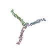


 PDBj
PDBj
 Assembly
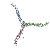

 Sample preparation
 / Beamline: 8.3.1 / Wavelength: 1.017 Å
 Processing