 ムービー
ムービー コントローラー


+ データを開く

- 基本情報
基本情報
| 登録情報 | データベース: EMDB / ID: EMD-2158 | |||||||||
|---|---|---|---|---|---|---|---|---|---|---|
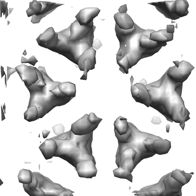
| タイトル | Bacterial chemoreceptor arrays are hexagonally packed trimers of receptor dimers networked by rings of kinase and coupling proteins | |||||||||
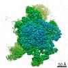
 マップデータ マップデータ | 3-fold average of wild-type E.coli (strain RP437) Chemoreceptor array sub-volumes (composed of 6 trimers-of receptor dimers) | |||||||||
 試料 試料 |
| |||||||||
 キーワード キーワード | chemotaxis / signal transduction / two-component systems | |||||||||
| 生物種 |  | |||||||||
| 手法 | サブトモグラム平均法 / クライオ電子顕微鏡法 | |||||||||
 データ登録者 データ登録者 | Briegel A / Li X / Bilwes AM / Hughes KT / Jensen GJ / Crane BR | |||||||||
 引用 引用 | ジャーナル: Proc Natl Acad Sci U S A / 年: 2012 タイトル: Bacterial chemoreceptor arrays are hexagonally packed trimers of receptor dimers networked by rings of kinase and coupling proteins. 著者: Ariane Briegel / Xiaoxiao Li / Alexandrine M Bilwes / Kelly T Hughes / Grant J Jensen / Brian R Crane /  要旨: Chemoreceptor arrays are supramolecular transmembrane machines of unknown structure that allow bacteria to sense their surroundings and respond by chemotaxis. We have combined X-ray crystallography ...Chemoreceptor arrays are supramolecular transmembrane machines of unknown structure that allow bacteria to sense their surroundings and respond by chemotaxis. We have combined X-ray crystallography of purified proteins with electron cryotomography of native arrays inside cells to reveal the arrangement of the component transmembrane receptors, histidine kinases (CheA) and CheW coupling proteins. Trimers of receptor dimers lie at the vertices of a hexagonal lattice in a "two-facing-two" configuration surrounding a ring of alternating CheA regulatory domains (P5) and CheW couplers. Whereas the CheA kinase domains (P4) project downward below the ring, the CheA dimerization domains (P3) link neighboring rings to form an extended, stable array. This highly interconnected protein architecture underlies the remarkable sensitivity and cooperative nature of transmembrane signaling in bacterial chemotaxis. | |||||||||
| 履歴 |
|
- 構造の表示
構造の表示
| ムービー |
 ムービービューア ムービービューア |
|---|---|
| 構造ビューア | EMマップ: SurfViewMolmilJmol/JSmol |
| 添付画像 |
- ダウンロードとリンク
-EMDBアーカイブ
| マップデータ | emd_2158.map.gz | 223.1 KB | EMDBマップデータ形式 | |
|---|---|---|---|---|
| ヘッダ (付随情報) | emd-2158-v30.xmlemd-2158.xml | 11 KB 11 KB | 表示 表示 | EMDBヘッダ |
| 画像 |  EMD-2158.png EMD-2158.png | 74.8 KB | ||
| アーカイブディレクトリ |  http://ftp.pdbj.org/pub/emdb/structures/EMD-2158ftp://ftp.pdbj.org/pub/emdb/structures/EMD-2158 http://ftp.pdbj.org/pub/emdb/structures/EMD-2158ftp://ftp.pdbj.org/pub/emdb/structures/EMD-2158 | HTTPS FTP |
-検証レポート
| 文書・要旨 | emd_2158_validation.pdf.gz | 208.7 KB | 表示 | EMDB検証レポート |
|---|---|---|---|---|
| 文書・詳細版 | emd_2158_full_validation.pdf.gz | 207.8 KB | 表示 | |
| XML形式データ | emd_2158_validation.xml.gz | 4.7 KB | 表示 | |
| アーカイブディレクトリ | https://ftp.pdbj.org/pub/emdb/validation_reports/EMD-2158ftp://ftp.pdbj.org/pub/emdb/validation_reports/EMD-2158 | HTTPS FTP |
-関連構造データ

-リンク
| EMDBのページ | EMDB (EBI/PDBe) / EMDataResource |
|---|
-マップ
| ファイル | ダウンロード / ファイル: emd_2158.map.gz / 形式: CCP4 / 大きさ: 297.9 KB / タイプ: IMAGE STORED AS FLOATING POINT NUMBER (4 BYTES) | ||||||||||||||||||||||||||||||||||||||||||||||||||||||||||||||||||||
|---|---|---|---|---|---|---|---|---|---|---|---|---|---|---|---|---|---|---|---|---|---|---|---|---|---|---|---|---|---|---|---|---|---|---|---|---|---|---|---|---|---|---|---|---|---|---|---|---|---|---|---|---|---|---|---|---|---|---|---|---|---|---|---|---|---|---|---|---|---|
| 注釈 | 3-fold average of wild-type E.coli (strain RP437) Chemoreceptor array sub-volumes (composed of 6 trimers-of receptor dimers) | ||||||||||||||||||||||||||||||||||||||||||||||||||||||||||||||||||||
| 投影像・断面図 | 画像のコントロール
画像は Spider により作成 これらの図は立方格子座標系で作成されたものです | ||||||||||||||||||||||||||||||||||||||||||||||||||||||||||||||||||||
| ボクセルのサイズ | X=Y=Z: 6.48 Å | ||||||||||||||||||||||||||||||||||||||||||||||||||||||||||||||||||||
| 密度 |
| ||||||||||||||||||||||||||||||||||||||||||||||||||||||||||||||||||||
| 対称性 | 空間群: 1 | ||||||||||||||||||||||||||||||||||||||||||||||||||||||||||||||||||||
| 詳細 | EMDB XML:
CCP4マップ ヘッダ情報:
| ||||||||||||||||||||||||||||||||||||||||||||||||||||||||||||||||||||
 Z (Sec.)
 Y (Row.)
 X (Col.)


-添付データ
- 試料の構成要素
-全体 : Bacterial chemoreceptor arrays
| 全体 | 名称: Bacterial chemoreceptor arrays |
|---|---|
| 要素 |
|
-超分子 #1000: Bacterial chemoreceptor arrays
| 超分子 | 名称: Bacterial chemoreceptor arrays / タイプ: sample / ID: 1000 集合状態: Trimers of receptor dimers lie at the vertices of a hexagonal lattice in a two-facing-two configuration surrounding a ring of alternating CheA regulatory domains (P5) and CheW couplers Number unique components: 3 |
|---|
-分子 #1: cheA
| 分子 | 名称: cheA / タイプ: protein_or_peptide / ID: 1 / 組換発現: No / データベース: NCBI |
|---|---|
| 由来(天然) | 生物種: |
-分子 #2: cheW
| 分子 | 名称: cheW / タイプ: protein_or_peptide / ID: 2 / 組換発現: No / データベース: NCBI |
|---|---|
| 由来(天然) | 生物種: |
-分子 #3: Chemoreceptors
| 分子 | 名称: Chemoreceptors / タイプ: protein_or_peptide / ID: 3 / 組換発現: No / データベース: NCBI |
|---|---|
| 由来(天然) | 生物種: |
-実験情報
-構造解析
| 手法 | クライオ電子顕微鏡法 |
|---|---|
 解析 解析 | サブトモグラム平均法 |
-試料調製
| 緩衝液 | 詳細: Tryptone Broth (10 g Tryptone, 5 g NaCl per liter). Cells were grown to exponential phase and incubated with penicillin for 1 hour prior to freezing |
|---|---|
| グリッド | 詳細: R 2/2 copper/Rhodium grids, glow discharged |
| 凍結 | 凍結剤: ETHANE / チャンバー内湿度: 100 % / 装置: FEI VITROBOT MARK III |
- 電子顕微鏡法
| 顕微鏡 | FEI POLARA 300 |
|---|---|
| 温度 | 平均: 77 K |
| 特殊光学系 | エネルギーフィルター - 名称: Gif 2002 hybrid (Gatan) エネルギーフィルター - エネルギー下限: 0.0 eV エネルギーフィルター - エネルギー上限: 20.0 eV |
| 日付 | 2008年1月15日 |
| 撮影 | カテゴリ: CCD フィルム・検出器のモデル: GATAN ULTRASCAN 4000 (4k x 4k) 平均電子線量: 150 e/Å2 |
| 電子線 | 加速電圧: 300 kV / 電子線源:  FIELD EMISSION GUN FIELD EMISSION GUN |
| 電子光学系 | 照射モード: FLOOD BEAM / 撮影モード: BRIGHT FIELD / Cs: 2.2 mm / 最小 デフォーカス(公称値): 10.0 µm / 倍率(公称値): 34000 |
| 試料ステージ | 試料ホルダーモデル: OTHER / Tilt series - Axis1 - Min angle: -70 ° / Tilt series - Axis1 - Max angle: 58 ° |
| 実験機器 |  モデル: Tecnai Polara / 画像提供: FEI Company |
-画像解析
| 詳細 | CTF correction in imod. Average number of tilts used in the 3D reconstructions: 130. Average tomographic tilt angle increment: 1. |
|---|---|
| 最終 再構成 | ソフトウェア - 名称: imod |
| CTF補正 | 詳細: imod |
-原子モデル構築 1
| 初期モデル | PDB ID: Chain - #0 - Chain ID: A / Chain - #1 - Chain ID: B / Chain - #2 - Chain ID: C / Chain - #3 - Chain ID: D |
|---|---|
| 詳細 | Protocol: rigid body |
| 精密化 | 空間: REAL / プロトコル: RIGID BODY FIT |

