National Institutes of Health/National Institute of Neurological Disorders and Stroke (NIH/NINDS)
NS092695
米国
引用
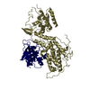
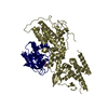
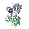
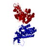
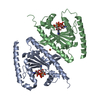
ジャーナル: Cell Rep / 年: 2020 タイトル: Structures of Gα Proteins in Complex with Their Chaperone Reveal Quality Control Mechanisms. 著者: Alpay Burak Seven / Daniel Hilger / Makaía M Papasergi-Scott / Li Zhang / Qianhui Qu / Brian K Kobilka / Gregory G Tall / Georgios Skiniotis / 要旨: Many chaperones promote nascent polypeptide folding followed by substrate release through ATP-dependent conformational changes. Here we show cryoEM structures of Gα subunit folding intermediates in ...Many chaperones promote nascent polypeptide folding followed by substrate release through ATP-dependent conformational changes. Here we show cryoEM structures of Gα subunit folding intermediates in complex with full-length Ric-8A, a unique chaperone-client system in which substrate release is facilitated by guanine nucleotide binding to the client G protein. The structures of Ric-8A-Gα and Ric-8A-Gα complexes reveal that the chaperone employs its extended C-terminal region to cradle the Ras-like domain of Gα, positioning the Ras core in contact with the Ric-8A core while engaging its switch2 nucleotide binding region. The C-terminal α5 helix of Gα is held away from the Ras-like domain through Ric-8A core domain interactions, which critically depend on recognition of the Gα C terminus by the chaperone. The structures, complemented with biochemical and cellular chaperoning data, support a folding quality control mechanism that ensures proper formation of the C-terminal α5 helix before allowing GTP-gated release of Gα from Ric-8A.
 ムービー
ムービー コントローラー


 データを開く
 基本情報
基本情報 マップデータ
 試料
試料 キーワード
キーワード 機能・相同性情報
 Homo sapiens (ヒト)
 データ登録者
 米国, 1件
  引用
引用 構造の表示
構造の表示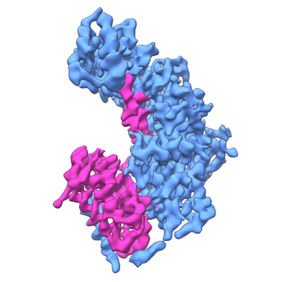
 ダウンロードとリンク
 emd_21387.png
 http://ftp.pdbj.org/pub/emdb/structures/EMD-21387


 Z (Sec.)
 Y (Row.)
 X (Col.)


 試料の構成要素
 Trichoplusia ni (イラクサキンウワバ)
 解析
解析 電子顕微鏡法
 FIELD EMISSION GUN
