- EMDB-21250: Cryo-EM structure of the C-terminal half of the Parkinson's Disea... -
+
Open data
ID or keywords:
Loading...
-
Basic information
Entry
Database: EMDB / ID: EMD-21250
Title
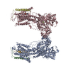
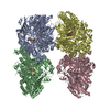
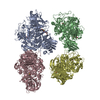
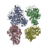
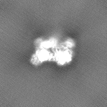
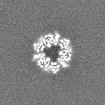
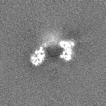
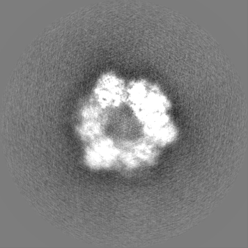
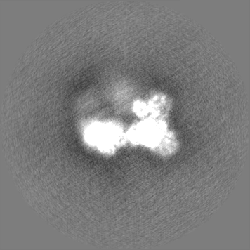
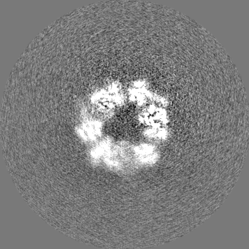
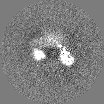
Cryo-EM structure of the C-terminal half of the Parkinson's Disease-linked protein Leucine Rich Repeat Kinase 2 (LRRK2)
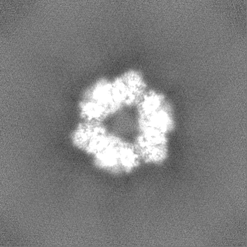
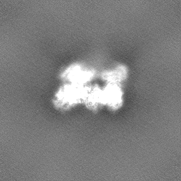
Map data
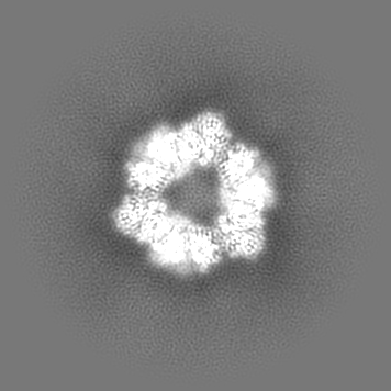
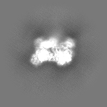
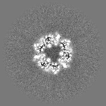
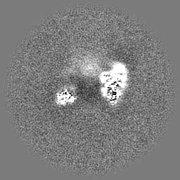
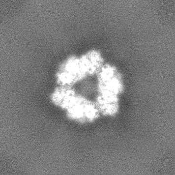
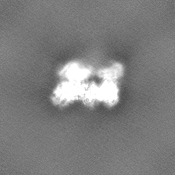
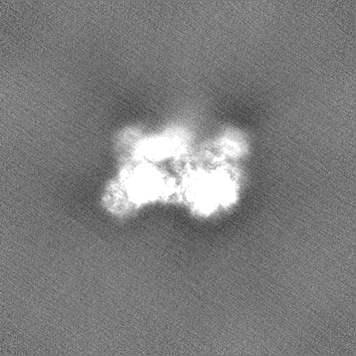
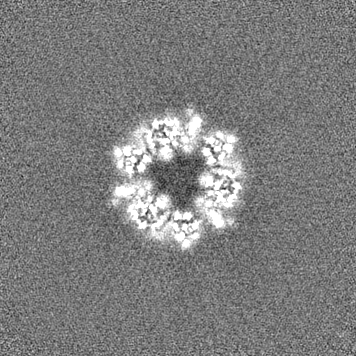
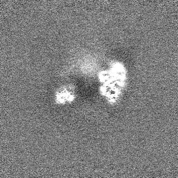
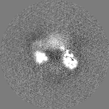
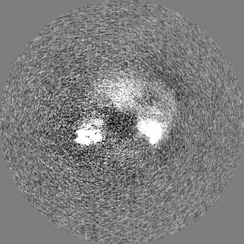
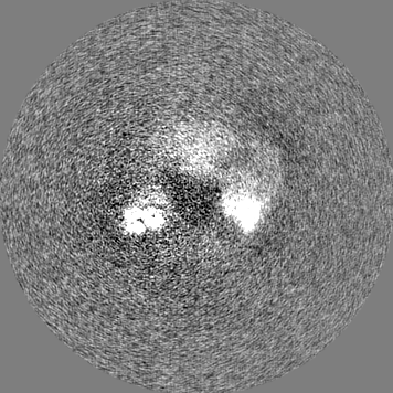
3.8A signal subtracted locally filtered cryo-EM map of C-terminal half of Leucine Rich Repeat Kinase 2 (LRRK2)
Sample
Complex: The C-terminal half of the Leucine Rich Repeat Kinase 2 (LRRK2) protein.
Protein or peptide: Leucine-rich repeat serine/threonine-protein kinase 2
Ligand: GUANOSINE-5'-DIPHOSPHATE
Ligand: MAGNESIUM ION
Keywords
Kinase / GTPase / SIGNALING PROTEIN
Function / homology
Function and homology information
caveola neck / negative regulation of protein processing involved in protein targeting to mitochondrion / Wnt signalosome assembly / beta-catenin destruction complex binding / regulation of branching morphogenesis of a nerve / regulation of kidney size / regulation of cell projection organization / tangential migration from the subventricular zone to the olfactory bulb / protein localization to endoplasmic reticulum exit site / GTP-dependent protein kinase activity ...caveola neck / negative regulation of protein processing involved in protein targeting to mitochondrion / Wnt signalosome assembly / beta-catenin destruction complex binding / regulation of branching morphogenesis of a nerve / regulation of kidney size / regulation of cell projection organization / tangential migration from the subventricular zone to the olfactory bulb / protein localization to endoplasmic reticulum exit site / GTP-dependent protein kinase activity / regulation of SNARE complex assembly / regulation of neuroblast proliferation / regulation of ER to Golgi vesicle-mediated transport / peroxidase inhibitor activity / negative regulation of late endosome to lysosome transport / regulation of mitochondrial depolarization / negative regulation of protein targeting to mitochondrion / positive regulation of dopamine receptor signaling pathway / regulation of synaptic vesicle transport / amphisome / regulation of CAMKK-AMPK signaling cascade / regulation of lysosomal lumen pH / negative regulation of GTPase activity / co-receptor binding / regulation of dopamine receptor signaling pathway / mitochondrion localization / regulation of neuron maturation / positive regulation of microglial cell activation / regulation of retrograde transport, endosome to Golgi / positive regulation of synaptic vesicle endocytosis / negative regulation of autophagosome assembly / cytoplasmic side of mitochondrial outer membrane / negative regulation of excitatory postsynaptic potential / JUN kinase kinase kinase activity / olfactory bulb development / neuron projection arborization / multivesicular body, internal vesicle / striatum development / regulation of dendritic spine morphogenesis / protein localization to mitochondrion / cellular response to dopamine / positive regulation of mitochondrial outer membrane permeabilization involved in apoptotic signaling pathway / endoplasmic reticulum organization / positive regulation of protein autoubiquitination / Wnt signalosome / positive regulation of programmed cell death / negative regulation of protein processing / GTP metabolic process / regulation of canonical Wnt signaling pathway / syntaxin-1 binding / regulation of reactive oxygen species metabolic process / lysosome organization / Golgi-associated vesicle / clathrin binding / protein kinase A binding / PTK6 promotes HIF1A stabilization / regulation of locomotion / negative regulation of macroautophagy / neuromuscular junction development / regulation of cAMP/PKA signal transduction / regulation of mitochondrial fission / Golgi organization / regulation of synaptic vesicle exocytosis / exploration behavior / microvillus / intracellular distribution of mitochondria / endoplasmic reticulum exit site / locomotory exploration behavior / autolysosome / negative regulation of Notch signaling pathway / MAP kinase kinase kinase activity / regulation of synaptic vesicle endocytosis / canonical Wnt signaling pathway / regulation of synaptic transmission, glutamatergic / negative regulation of endoplasmic reticulum stress-induced intrinsic apoptotic signaling pathway / presynaptic cytosol / Rho protein signal transduction / phagocytic vesicle / neuron projection morphogenesis / JNK cascade / cellular response to manganese ion / positive regulation of autophagy / tubulin binding / dendrite cytoplasm / GTPase activator activity / cellular response to starvation / positive regulation of protein ubiquitination / SNARE binding / determination of adult lifespan / cellular response to reactive oxygen species / regulation of membrane potential / excitatory postsynaptic potential / mitochondrion organization / trans-Golgi network / calcium-mediated signaling / mitochondrial membrane / regulation of protein stability / small GTPase binding / autophagy / endocytosis Similarity search - Function
: / : / : / LRRK2 ARM repeat / LRRK2 ANK repeat / LRRK2 beta propeller / : / C-terminal of Roc, COR-B domain / C-terminal of Roc (COR) domain / C-terminal of Roc, COR-A domain ...: / : / : / LRRK2 ARM repeat / LRRK2 ANK repeat / LRRK2 beta propeller / : / C-terminal of Roc, COR-B domain / C-terminal of Roc (COR) domain / C-terminal of Roc, COR-A domain / Ras of Complex, Roc, domain of DAPkinase / Roc domain profile. / Roc domain / Leucine-rich repeats, bacterial type / Leucine rich repeat / Leucine-rich repeat, typical subtype / Leucine-rich repeats, typical (most populated) subfamily / Leucine-rich repeat profile. / Leucine-rich repeat / Rab subfamily of small GTPases / Leucine-rich repeat domain superfamily / Ankyrin repeat-containing domain superfamily / Armadillo-like helical / Small GTP-binding protein domain / Armadillo-type fold / Serine/threonine-protein kinase, active site / Serine/Threonine protein kinases active-site signature. / WD40-repeat-containing domain superfamily / Protein kinase domain / Serine/Threonine protein kinases, catalytic domain / WD40/YVTN repeat-like-containing domain superfamily / Protein kinase, ATP binding site / Protein kinases ATP-binding region signature. / Protein kinase domain profile. / Protein kinase domain / Protein kinase-like domain superfamily / P-loop containing nucleoside triphosphate hydrolase Similarity search - Domain/homology
National Institutes of Health/National Institute of General Medical Sciences (NIH/NIGMS)
R01GM107214
United States
Citation
Journal: Nature / Year: 2020 Title: Structure of LRRK2 in Parkinson's disease and model for microtubule interaction. Authors: C K Deniston / J Salogiannis / S Mathea / D M Snead / I Lahiri / M Matyszewski / O Donosa / R Watanabe / J Böhning / A K Shiau / S Knapp / E Villa / S L Reck-Peterson / A E Leschziner / Abstract: Leucine-rich repeat kinase 2 (LRRK2) is the most commonly mutated gene in familial Parkinson's disease and is also linked to its idiopathic form. LRRK2 has been proposed to function in membrane ...Leucine-rich repeat kinase 2 (LRRK2) is the most commonly mutated gene in familial Parkinson's disease and is also linked to its idiopathic form. LRRK2 has been proposed to function in membrane trafficking and colocalizes with microtubules. Despite the fundamental importance of LRRK2 for understanding and treating Parkinson's disease, structural information on the enzyme is limited. Here we report the structure of the catalytic half of LRRK2, and an atomic model of microtubule-associated LRRK2 built using a reported cryo-electron tomography in situ structure. We propose that the conformation of the LRRK2 kinase domain regulates its interactions with microtubules, with a closed conformation favouring oligomerization on microtubules. We show that the catalytic half of LRRK2 is sufficient for filament formation and blocks the motility of the microtubule-based motors kinesin 1 and cytoplasmic dynein 1 in vitro. Kinase inhibitors that stabilize an open conformation relieve this interference and reduce the formation of LRRK2 filaments in cells, whereas inhibitors that stabilize a closed conformation do not. Our findings suggest that LRRK2 can act as a roadblock for microtubule-based motors and have implications for the design of therapeutic LRRK2 kinase inhibitors.
History
Deposition
Jan 29, 2020
-
Header (metadata) release
Aug 26, 2020
-
Map release
Aug 26, 2020
-
Update
Nov 20, 2024
-
Current status
Nov 20, 2024
Processing site: RCSB / Status: Released
-
Structure visualization
Movie
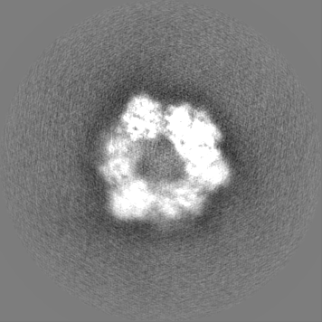
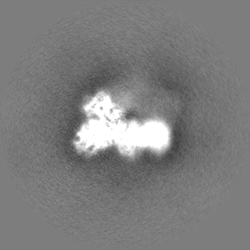
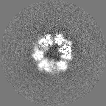
Surface view with section colored by density value
Cryogen name: ETHANE / Chamber humidity: 100 % / Chamber temperature: 277 K / Instrument: FEI VITROBOT MARK II
Details
4uM concentration
-
Electron microscopy
Microscope
FEI TITAN KRIOS
Specialist optics
Energy filter - Name: GIF 2002
Image recording
Film or detector model: GATAN K2 SUMMIT (4k x 4k) / Detector mode: COUNTING / Number grids imaged: 1 / Number real images: 3826 / Average exposure time: 8.0 sec. / Average electron dose: 6.65 e/Å2
Electron beam
Acceleration voltage: 300 kV / Electron source: FIELD EMISSION GUN
Type of model: OTHER Details: Generated initial models from ab initio refinement in Cryosparc.
Final reconstruction
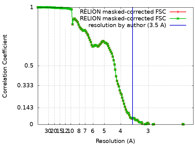
Applied symmetry - Point group: C3 (3 fold cyclic) / Resolution.type: BY AUTHOR / Resolution: 3.5 Å / Resolution method: FSC 0.143 CUT-OFF / Software:
Name
details
RELION (ver. 3)
C1
cryoSPARC (ver. 2)
C3
Details: For the signal subtracted map, 105,787 (tripled) particles went into the final map that achieved 3.8A resolution. Number images used: 70953
Initial angle assignment
Type: MAXIMUM LIKELIHOOD / Software: (Name: RELION (ver. 3), cryoSPARC (ver. 2)) Details: Signal subtracted map was generated in Relion3. The C3 map however was created in Cryosparc 2.
Final angle assignment
Type: MAXIMUM LIKELIHOOD / Software: (Name: RELION (ver. 3), cryoSPARC (ver. 2)) Details: Signal subtracted map was generated in Relion3. The C3 map however was created in Cryosparc 2.
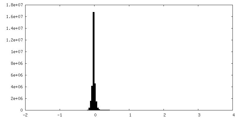
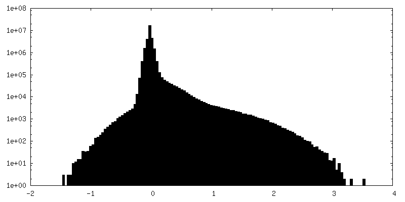
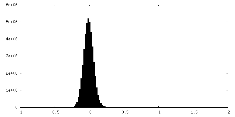
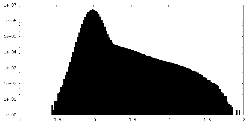
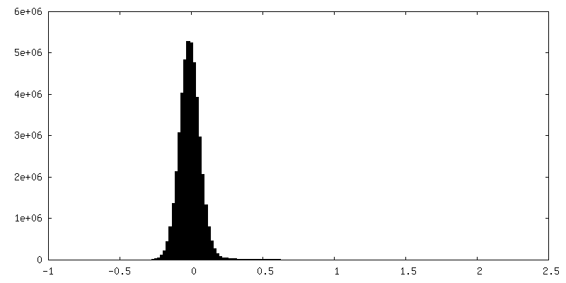
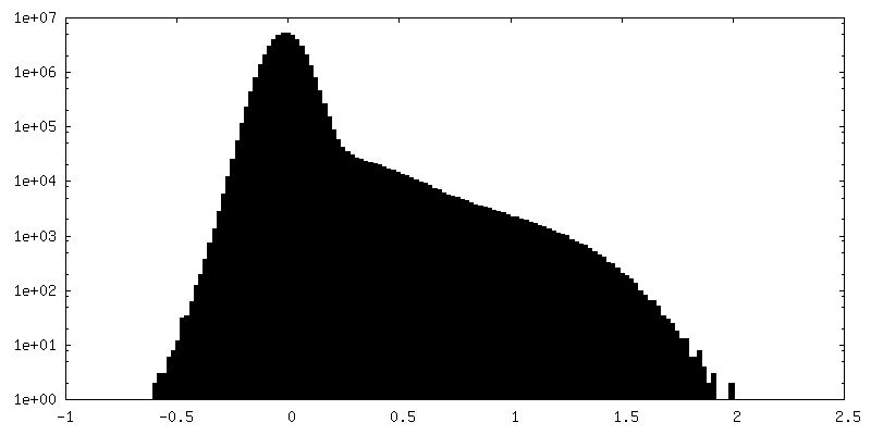
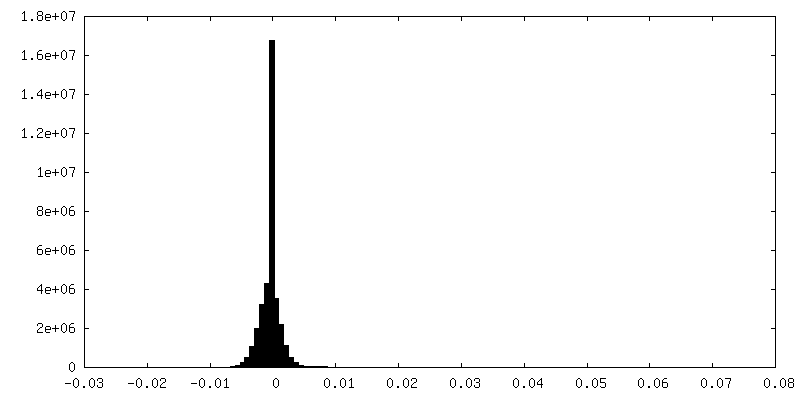
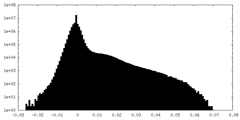
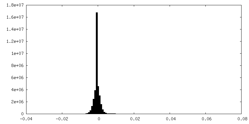
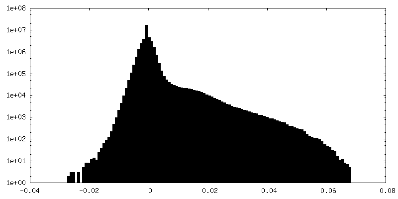
FSC plot (resolution estimation)
+
About Yorodumi
-
News
-
Feb 9, 2022. New format data for meta-information of EMDB entries
New format data for meta-information of EMDB entries
Version 3 of the EMDB header file is now the official format.
The previous official version 1.9 will be removed from the archive.
In the structure databanks used in Yorodumi, some data are registered as the other names, "COVID-19 virus" and "2019-nCoV". Here are the details of the virus and the list of structure data.
Jan 31, 2019. EMDB accession codes are about to change! (news from PDBe EMDB page)
EMDB accession codes are about to change! (news from PDBe EMDB page)
The allocation of 4 digits for EMDB accession codes will soon come to an end. Whilst these codes will remain in use, new EMDB accession codes will include an additional digit and will expand incrementally as the available range of codes is exhausted. The current 4-digit format prefixed with “EMD-” (i.e. EMD-XXXX) will advance to a 5-digit format (i.e. EMD-XXXXX), and so on. It is currently estimated that the 4-digit codes will be depleted around Spring 2019, at which point the 5-digit format will come into force.
The EM Navigator/Yorodumi systems omit the EMD- prefix.
Related info.:Q: What is EMD? / ID/Accession-code notation in Yorodumi/EM Navigator
Yorodumi is a browser for structure data from EMDB, PDB, SASBDB, etc.
This page is also the successor to EM Navigator detail page, and also detail information page/front-end page for Omokage search.
The word "yorodu" (or yorozu) is an old Japanese word meaning "ten thousand". "mi" (miru) is to see.
Related info.:EMDB / PDB / SASBDB / Comparison of 3 databanks / Yorodumi Search / Aug 31, 2016. New EM Navigator & Yorodumi / Yorodumi Papers / Jmol/JSmol / Function and homology information / Changes in new EM Navigator and Yorodumi
 Movie
Movie Controller

 Yorodumi
 Open data
 Basic information
 Map data
 Sample
 Keywords
 Function and homology information
 Homo sapiens (human)
 Authors
 United States, 3 items
  Citation


 Structure visualization
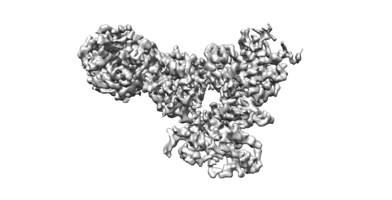
 Downloads & links
 emd_21250.png
 http://ftp.pdbj.org/pub/emdb/structures/EMD-21250


 Z (Sec.)
 Y (Row.)
 X (Col.)


 Sample components
 Spodoptera frugiperda (fall armyworm)
 Processing
 Electron microscopy
 FIELD EMISSION GUN