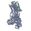
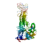
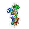
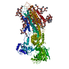
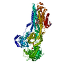
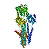
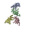
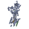
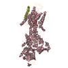
- PDB-3b8e: Crystal structure of the sodium-potassium pump -
+
Open data
ID or keywords:
Loading...
-
Basic information
Entry
Database: PDB / ID: 3b8e
Title
Crystal structure of the sodium-potassium pump
Components
(Sodium/potassium-transporting ATPase subunit ...) x 2
Na+/K+ ATPase gamma subunit transcript variant a
Keywords
HYDROLASE/TRANSPORT PROTEIN / Na+ / K+-ATPASE / P-TYPE ATPASE / CATION PUMP / MEMBRANE PROTEIN / HYDROLASE / ATP-BINDING / CALCIUM TRANSPORT / ION TRANSPORT / MEMBRANE POTENTIAL / PHOSPHORYLATION / Magnesium / Metal-binding / Nucleotide-binding / Potassium / Potassium transport / Sodium / Sodium transport / Sodium/potassium transport / Transmembrane / Glycoprotein / Signal-anchor / HYDROLASE-TRANSPORT PROTEIN COMPLEX
Function / homology
Function and homology information
: / Ion homeostasis / Ion transport by P-type ATPases / Na+/K+-exchanging ATPase / regulation of monoatomic ion transport / positive regulation of sodium ion export across plasma membrane / positive regulation of potassium ion import across plasma membrane / P-type sodium:potassium-exchanging transporter activity / sodium ion binding / sodium:potassium-exchanging ATPase complex ...: / Ion homeostasis / Ion transport by P-type ATPases / Na+/K+-exchanging ATPase / regulation of monoatomic ion transport / positive regulation of sodium ion export across plasma membrane / positive regulation of potassium ion import across plasma membrane / P-type sodium:potassium-exchanging transporter activity / sodium ion binding / sodium:potassium-exchanging ATPase complex / membrane repolarization / establishment or maintenance of transmembrane electrochemical gradient / sodium ion export across plasma membrane / regulation of calcium ion transmembrane transport / positive regulation of potassium ion transmembrane transport / ion channel regulator activity / intracellular sodium ion homeostasis / relaxation of cardiac muscle / regulation of cardiac muscle contraction by calcium ion signaling / positive regulation of sodium ion transmembrane transport / organelle membrane / potassium ion binding / potassium ion import across plasma membrane / ATPase activator activity / intracellular potassium ion homeostasis / intercalated disc / lateral plasma membrane / transporter activator activity / sperm flagellum / ATP metabolic process / regulation of sodium ion transport / cardiac muscle contraction / T-tubule / proton transmembrane transport / protein localization to plasma membrane / sarcolemma / transmembrane transport / intracellular calcium ion homeostasis / melanosome / ATPase binding / regulation of gene expression / basolateral plasma membrane / protein-macromolecule adaptor activity / cell adhesion / apical plasma membrane / protein stabilization / innate immune response / axon / protein kinase binding / ATP hydrolysis activity / ATP binding / membrane / plasma membrane Similarity search - Function
Mass: 790.145 Da / Num. of mol.: 2 / Source method: obtained synthetically / Formula: C44H88NO8P / Comment: phospholipid*YM
-
Details
Nonpolymer details
THE AUTHORS ONLY SEE DENSITY FOR THE PHOSPHATIDYLCHOLINE LIPID HEAD GROUP AND THEY DO NOT HAVE ANY ...THE AUTHORS ONLY SEE DENSITY FOR THE PHOSPHATIDYLCHOLINE LIPID HEAD GROUP AND THEY DO NOT HAVE ANY EXPERIMENTAL EVIDENCE FOR PC1.
-
Experimental details
-
Experiment
Experiment
Method: X-RAY DIFFRACTION / Number of used crystals: 1
-
Sample preparation
Crystal
Density Matthews: 6.35 Å3/Da / Density % sol: 80.63 %
Resolution: 3.5→3.6 Å / Redundancy: 14 % / Rmerge(I) obs: 1.2 / Mean I/σ(I) obs: 2.56 / Num. unique all: 6208 / % possible all: 100
-
Processing
Software
Name
Version
Classification
SHARP
phasing
CNS
1.2
refinement
XDS
datareduction
XSCALE
datascaling
Refinement
Method to determine structure: MIR / Resolution: 3.5→20 Å / Isotropic thermal model: GROUPED B-factors / Stereochemistry target values: Engh & Huber
Rfactor
Num. reflection
% reflection
Selection details
Rfree
0.3129
1541
-
RANDOM
Rwork
0.2774
-
-
-
all
-
77267
-
-
obs
-
76989
99.6 %
-
Displacement parameters
Biso mean: 104 Å2
Baniso -1
Baniso -2
Baniso -3
1-
3.508 Å2
0 Å2
0 Å2
2-
-
17.739 Å2
0 Å2
3-
-
-
-21.247 Å2
Refine analyze
Free
Obs
Luzzati coordinate error
0.6 Å
0.54 Å
Luzzati d res low
-
5 Å
Luzzati sigma a
1.1 Å
1.15 Å
Refinement step
Cycle: LAST / Resolution: 3.5→20 Å
Protein
Nucleic acid
Ligand
Solvent
Total
Num. atoms
16676
0
42
0
16718
Refine LS restraints
Refine-ID
Type
Dev ideal
X-RAY DIFFRACTION
c_bond_d
0.008002
X-RAY DIFFRACTION
c_angle_deg
1.51826
X-RAY DIFFRACTION
c_improper_angle_d
1.086
X-RAY DIFFRACTION
c_dihedral_angle_d
22.642
Refine LS restraints NCS
NCS model details: RESTRAINED NCS ON DOMAINS: A(resid 19:80 and 154:274 and 344:382 and 592:765), A(383:591), A(91:153), A(275:343 and 2003:2005), A(766:1016 and B and G) Rms dev Biso: 3 Å2 / Weight position: 500
LS refinement shell
Resolution: 3.5→3.54 Å
Rfactor
Num. reflection
Rfree
0.385
39
Rwork
0.4291
-
obs
-
2279
+
About Yorodumi
-
News
-
Feb 9, 2022. New format data for meta-information of EMDB entries
New format data for meta-information of EMDB entries
Version 3 of the EMDB header file is now the official format.
The previous official version 1.9 will be removed from the archive.
In the structure databanks used in Yorodumi, some data are registered as the other names, "COVID-19 virus" and "2019-nCoV". Here are the details of the virus and the list of structure data.
Jan 31, 2019. EMDB accession codes are about to change! (news from PDBe EMDB page)
EMDB accession codes are about to change! (news from PDBe EMDB page)
The allocation of 4 digits for EMDB accession codes will soon come to an end. Whilst these codes will remain in use, new EMDB accession codes will include an additional digit and will expand incrementally as the available range of codes is exhausted. The current 4-digit format prefixed with “EMD-” (i.e. EMD-XXXX) will advance to a 5-digit format (i.e. EMD-XXXXX), and so on. It is currently estimated that the 4-digit codes will be depleted around Spring 2019, at which point the 5-digit format will come into force.
The EM Navigator/Yorodumi systems omit the EMD- prefix.
Related info.:Q: What is EMD? / ID/Accession-code notation in Yorodumi/EM Navigator
Yorodumi is a browser for structure data from EMDB, PDB, SASBDB, etc.
This page is also the successor to EM Navigator detail page, and also detail information page/front-end page for Omokage search.
The word "yorodu" (or yorozu) is an old Japanese word meaning "ten thousand". "mi" (miru) is to see.
Related info.:EMDB / PDB / SASBDB / Comparison of 3 databanks / Yorodumi Search / Aug 31, 2016. New EM Navigator & Yorodumi / Yorodumi Papers / Jmol/JSmol / Function and homology information / Changes in new EM Navigator and Yorodumi
 Movie
Movie Controller


 Open data
 Basic information
 Components
 Keywords
 Function and homology information
 X-RAY DIFFRACTION /
  Authors
 Citation
 Structure visualization
 Downloads & links
 Other downloads
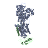
 PDBj
PDBj


 Assembly
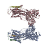


 Mass: 24.305 Da / Num. of mol.: 2 / Source method: obtained synthetically / Formula: Mg
 Mass: 85.468 Da / Num. of mol.: 6 / Source method: obtained synthetically / Formula: Rb
 Mass: 100.299 Da / Num. of mol.: 2 / Source method: obtained synthetically / Formula: F4Mg
 Mass: 790.145 Da / Num. of mol.: 2 / Source method: obtained synthetically / Formula: C44H88NO8P / Comment: phospholipid*YM
 Sample preparation
 / Beamline: X06SA / Wavelength: 1.078, 1.073, 0.81
 Processing