National Institutes of Health/National Institute of Neurological Disorders and Stroke (NIH/NINDS)
NS042169
United States
Citation
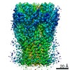
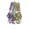
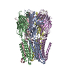
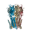
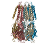
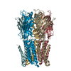
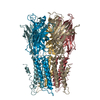
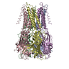
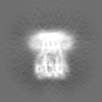
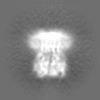
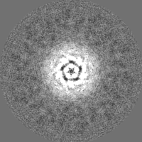
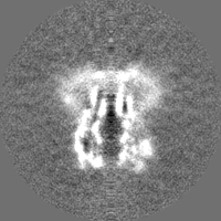
Journal: Proc Natl Acad Sci U S A / Year: 2020 Title: Cryo-EM structures of a lipid-sensitive pentameric ligand-gated ion channel embedded in a phosphatidylcholine-only bilayer. Authors: Pramod Kumar / Yuhang Wang / Zhening Zhang / Zhiyu Zhao / Gisela D Cymes / Emad Tajkhorshid / Claudio Grosman / Abstract: The lipid dependence of the nicotinic acetylcholine receptor from the electric organ has long been recognized, and one of the most consistent experimental observations is that, when reconstituted in ...The lipid dependence of the nicotinic acetylcholine receptor from the electric organ has long been recognized, and one of the most consistent experimental observations is that, when reconstituted in membranes formed by zwitterionic phospholipids alone, exposure to agonist fails to elicit ion-flux activity. More recently, it has been suggested that the bacterial homolog ELIC ( ligand-gated ion channel) has a similar lipid sensitivity. As a first step toward the elucidation of the structural basis of this phenomenon, we solved the structures of ELIC embedded in palmitoyl-oleoyl-phosphatidylcholine- (POPC-) only nanodiscs in both the unliganded (4.1-Å resolution) and agonist-bound (3.3 Å) states using single-particle cryoelectron microscopy. Comparison of the two structural models revealed that the largest differences occur at the level of loop C-at the agonist-binding sites-and the loops at the interface between the extracellular and transmembrane domains (ECD and TMD, respectively). On the other hand, the transmembrane pore is occluded in a remarkably similar manner in both structures. A straightforward interpretation of these findings is that POPC-only membranes frustrate the ECD-TMD coupling in such a way that the "conformational wave" of liganded-receptor gating takes place in the ECD and the interfacial M2-M3 linker but fails to penetrate the membrane and propagate into the TMD. Furthermore, analysis of the structural models and molecular simulations suggested that the higher affinity for agonists characteristic of the open- and desensitized-channel conformations results, at least in part, from the tighter confinement of the ligand to its binding site; this limits the ligand's fluctuations, and thus delays its escape into bulk solvent.
History
Deposition
Nov 18, 2019
-
Header (metadata) release
Dec 4, 2019
-
Map release
Jan 15, 2020
-
Update
Mar 6, 2024
-
Current status
Mar 6, 2024
Processing site: RCSB / Status: Released
-
Structure visualization
Movie
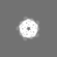
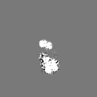
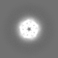
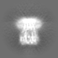
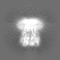
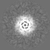
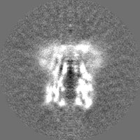
Surface view with section colored by density value
In the structure databanks used in Yorodumi, some data are registered as the other names, "COVID-19 virus" and "2019-nCoV". Here are the details of the virus and the list of structure data.
Jan 31, 2019. EMDB accession codes are about to change! (news from PDBe EMDB page)
EMDB accession codes are about to change! (news from PDBe EMDB page)
The allocation of 4 digits for EMDB accession codes will soon come to an end. Whilst these codes will remain in use, new EMDB accession codes will include an additional digit and will expand incrementally as the available range of codes is exhausted. The current 4-digit format prefixed with “EMD-” (i.e. EMD-XXXX) will advance to a 5-digit format (i.e. EMD-XXXXX), and so on. It is currently estimated that the 4-digit codes will be depleted around Spring 2019, at which point the 5-digit format will come into force.
The EM Navigator/Yorodumi systems omit the EMD- prefix.
Related info.:Q: What is EMD? / ID/Accession-code notation in Yorodumi/EM Navigator
Yorodumi is a browser for structure data from EMDB, PDB, SASBDB, etc.
This page is also the successor to EM Navigator detail page, and also detail information page/front-end page for Omokage search.
The word "yorodu" (or yorozu) is an old Japanese word meaning "ten thousand". "mi" (miru) is to see.
Related info.:EMDB / PDB / SASBDB / Comparison of 3 databanks / Yorodumi Search / Aug 31, 2016. New EM Navigator & Yorodumi / Yorodumi Papers / Jmol/JSmol / Function and homology information / Changes in new EM Navigator and Yorodumi
 Movie
Movie Controller


 Open data
 Basic information
 Map data
 Sample
 Keywords
 Function and homology information
 Dickeya dadantii 3937 (bacteria) /
  Authors
 United States, 1 items
  Citation
 Structure visualization
 Downloads & links
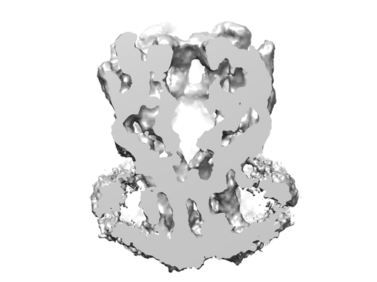 emd_20986.png
 http://ftp.pdbj.org/pub/emdb/structures/EMD-20986


 Z (Sec.)
 Y (Row.)
 X (Col.)


 Sample components
 Processing
 Electron microscopy
 FIELD EMISSION GUN
