 Movie
Movie Controller


+ Open data

- Basic information
| Entry | Database: PDB / ID: 6v0b | ||||||
|---|---|---|---|---|---|---|---|
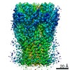
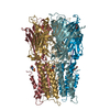
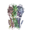
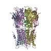
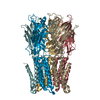
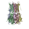
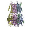
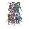
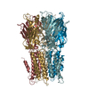
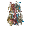
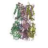
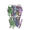
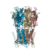
| Title | Unliganded ELIC in POPC-only nanodiscs. | ||||||
 Components Components | Gamma-aminobutyric-acid receptor subunit beta-1 | ||||||
 Keywords Keywords | MEMBRANE PROTEIN / Pentameric Ligand-gated Ion Channels / POPC / Nanodisc / Cys-loop receptors | ||||||
| Function / homology |  Function and homology information Function and homology informationextracellular ligand-gated monoatomic ion channel activity / transmembrane signaling receptor activity / identical protein binding / plasma membrane Similarity search - Function | ||||||
| Biological species |  Dickeya dadantii (bacteria) Dickeya dadantii (bacteria) | ||||||
| Method | ELECTRON MICROSCOPY / single particle reconstruction / cryo EM / Resolution: 4.1 Å | ||||||
 Authors Authors | Grosman, C. / Kumar, P. | ||||||
| Funding support |  United States, 1items United States, 1items
| ||||||
 Citation Citation | Journal: Proc Natl Acad Sci U S A / Year: 2020 Title: Cryo-EM structures of a lipid-sensitive pentameric ligand-gated ion channel embedded in a phosphatidylcholine-only bilayer. Authors: Pramod Kumar / Yuhang Wang / Zhening Zhang / Zhiyu Zhao / Gisela D Cymes / Emad Tajkhorshid / Claudio Grosman / Abstract: The lipid dependence of the nicotinic acetylcholine receptor from the electric organ has long been recognized, and one of the most consistent experimental observations is that, when reconstituted in ...The lipid dependence of the nicotinic acetylcholine receptor from the electric organ has long been recognized, and one of the most consistent experimental observations is that, when reconstituted in membranes formed by zwitterionic phospholipids alone, exposure to agonist fails to elicit ion-flux activity. More recently, it has been suggested that the bacterial homolog ELIC ( ligand-gated ion channel) has a similar lipid sensitivity. As a first step toward the elucidation of the structural basis of this phenomenon, we solved the structures of ELIC embedded in palmitoyl-oleoyl-phosphatidylcholine- (POPC-) only nanodiscs in both the unliganded (4.1-Å resolution) and agonist-bound (3.3 Å) states using single-particle cryoelectron microscopy. Comparison of the two structural models revealed that the largest differences occur at the level of loop C-at the agonist-binding sites-and the loops at the interface between the extracellular and transmembrane domains (ECD and TMD, respectively). On the other hand, the transmembrane pore is occluded in a remarkably similar manner in both structures. A straightforward interpretation of these findings is that POPC-only membranes frustrate the ECD-TMD coupling in such a way that the "conformational wave" of liganded-receptor gating takes place in the ECD and the interfacial M2-M3 linker but fails to penetrate the membrane and propagate into the TMD. Furthermore, analysis of the structural models and molecular simulations suggested that the higher affinity for agonists characteristic of the open- and desensitized-channel conformations results, at least in part, from the tighter confinement of the ligand to its binding site; this limits the ligand's fluctuations, and thus delays its escape into bulk solvent. | ||||||
| History |
|
- Structure visualization
| Movie |
Movie viewer |
|---|---|
| Structure viewer | Molecule: MolmilJmol/JSmol |
- Downloads & links
-Download
| PDBx/mmCIF format | 6v0b.cif.gz | 276.8 KB | Display | PDBx/mmCIF format |
|---|---|---|---|---|
| PDB format | pdb6v0b.ent.gz | 224.9 KB | Display | PDB format |
| PDBx/mmJSON format | 6v0b.json.gz | Tree view | PDBx/mmJSON format | |
| Others |  Other downloads Other downloads |
-Validation report
| Arichive directory | https://data.pdbj.org/pub/pdb/validation_reports/v0/6v0bftp://data.pdbj.org/pub/pdb/validation_reports/v0/6v0b | HTTPS FTP |
|---|
-Related structure data
| Related structure data |  20986MC  6v03C M: map data used to model this data C: citing same article ( |
|---|---|
| Similar structure data |

-Links
 PDBj
PDBj


- Assembly
| Deposited unit | 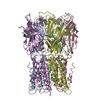
|
|---|---|
| 1 |
|
-Components
| #1: Protein | Mass: 36879.000 Da / Num. of mol.: 5 Source method: isolated from a genetically manipulated source Source: (gene. exp.) Dickeya dadantii (strain 3937) (bacteria)Strain: 3937 / Gene: Dda3937_00520 Production host: Strain (production host): BL21 / References: UniProt: E0SJQ4 |
|---|
-Experimental details
-Experiment
| Experiment | Method: ELECTRON MICROSCOPY |
|---|---|
| EM experiment | Aggregation state: PARTICLE / 3D reconstruction method: single particle reconstruction |
- Sample preparation
| Component | Name: Unliganded ELIC in POPC-only nanodiscs. / Type: COMPLEX / Entity ID: all / Source: RECOMBINANT | ||||||||||||||||||||
|---|---|---|---|---|---|---|---|---|---|---|---|---|---|---|---|---|---|---|---|---|---|
| Molecular weight | Experimental value: NO | ||||||||||||||||||||
| Source (natural) | Organism: Dickeya dadantii 3937 (bacteria) | ||||||||||||||||||||
| Source (recombinant) | Organism: | ||||||||||||||||||||
| Buffer solution | pH: 8 / Details: 150 mM NaCl and 10 mM sodium phosphate, pH 8.0. | ||||||||||||||||||||
| Buffer component |
| ||||||||||||||||||||
| Specimen | Conc.: 2 mg/ml / Embedding applied: NO / Shadowing applied: NO / Staining applied: NO / Vitrification applied: YES | ||||||||||||||||||||
| Specimen support | Grid material: GOLD / Grid type: Homemade | ||||||||||||||||||||
| Vitrification | Instrument: SPOTITON / Cryogen name: ETHANE |
- Electron microscopy imaging
| Experimental equipment |  Model: Titan Krios / Image courtesy: FEI Company |
|---|---|
| Microscopy | Model: FEI TITAN KRIOS |
| Electron gun | Electron source:  FIELD EMISSION GUN / Accelerating voltage: 300 kV / Illumination mode: SPOT SCAN FIELD EMISSION GUN / Accelerating voltage: 300 kV / Illumination mode: SPOT SCAN |
| Electron lens | Mode: BRIGHT FIELD / Cs: 2.7 mm |
| Image recording | Electron dose: 63.56 e/Å2 / Detector mode: COUNTING / Film or detector model: DIRECT ELECTRON DE-16 (4k x 4k) |
- Processing
| EM software | Name: RELION / Version: 2.1 / Category: 3D reconstruction | ||||||||||||||||||||||||
|---|---|---|---|---|---|---|---|---|---|---|---|---|---|---|---|---|---|---|---|---|---|---|---|---|---|
| CTF correction | Type: NONE | ||||||||||||||||||||||||
| Particle selection | Num. of particles selected: 289375 | ||||||||||||||||||||||||
| Symmetry | Point symmetry: C5 (5 fold cyclic) | ||||||||||||||||||||||||
| 3D reconstruction | Resolution: 4.1 Å / Resolution method: FSC 0.143 CUT-OFF / Num. of particles: 29207 / Symmetry type: POINT | ||||||||||||||||||||||||
| Atomic model building | Protocol: RIGID BODY FIT / Space: REAL | ||||||||||||||||||||||||
| Refinement | Stereochemistry target values: GeoStd + Monomer Library + CDL v1.2 | ||||||||||||||||||||||||
| Displacement parameters | Biso mean: 138.12 Å2 | ||||||||||||||||||||||||
| Refine LS restraints |
|
