Movie
Movie Controller

[English] 日本語
 Yorodumi
- EMDB-2065: Electron cryo-microscopy of R-peptide precursor of Moloney murine... -
+ Open data

- Basic information
| Entry | Database: EMDB / ID: EMD-2065 | |||||||||
|---|---|---|---|---|---|---|---|---|---|---|
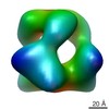
| Title | Electron cryo-microscopy of R-peptide precursor of Moloney murine leukemia virus Env in its isomerization arrested intermediate state | |||||||||
 Map data Map data | Reconstruction of the R-peptide precursor of Moloney murine leukemia virus Env in its isomerization arrested intermediate state | |||||||||
 Sample Sample |
| |||||||||
 Keywords Keywords | cryo-EM / Retrovirus / spike protein / maturation cleavage / R-peptide | |||||||||
| Biological species |  Moloney murine leukemia virus Moloney murine leukemia virus | |||||||||
| Method | single particle reconstruction / cryo EM / negative staining / Resolution: 22.0 Å | |||||||||
 Authors Authors | Loving R / Wu SR / Sjoberg M / Lindqvist B / Garoff H | |||||||||
 Citation Citation | Journal: Proc Natl Acad Sci U S A / Year: 2012 Title: Maturation cleavage of the murine leukemia virus Env precursor separates the transmembrane subunits to prime it for receptor triggering. Authors: Robin Löving / Shang-Rung Wu / Mathilda Sjöberg / Birgitta Lindqvist / Henrik Garoff /  Abstract: The Env protein of murine leukemia virus matures by two cleavage events. First, cellular furin separates the receptor binding surface (SU) subunit from the fusion-active transmembrane (TM) subunit ...The Env protein of murine leukemia virus matures by two cleavage events. First, cellular furin separates the receptor binding surface (SU) subunit from the fusion-active transmembrane (TM) subunit and then, in the newly assembled particle, the viral protease removes a 16-residue peptide, the R-peptide from the endodomain of the TM. Both cleavage events are required to prime the Env for receptor-triggered activation. Cryoelectron microscopy (cryo-EM) analyses have shown that the mature Env forms an open cage-like structure composed of three SU-TM complexes, where the TM subunits formed separated Env legs. Here we have studied the structure of the R-peptide precursor Env by cryo-EM. TM cleavage in Moloney murine leukemia virus was inhibited by amprenavir, and the Envs were solubilized in Triton X-100 and isolated by sedimentation in a sucrose gradient. We found that the legs of the R-peptide Env were held together by trimeric interactions at the very bottom of the Env. This suggested that the R-peptide ties the TM legs together and that this prevents the activation of the TM for fusion. The model was supported by further cryo-EM studies using an R-peptide Env mutant that was fusion-competent despite an uncleaved R-peptide. The Env legs of this mutant were found to be separated, like in the mature Env. This shows that it is the TM leg separation, normally caused by R-peptide cleavage, that primes the Env for receptor triggering. | |||||||||
| History |
|
- Structure visualization
| Movie |
 Movie viewer Movie viewer |
|---|---|
| Structure viewer | EM map: SurfViewMolmilJmol/JSmol |
| Supplemental images |

- Downloads & links
-EMDB archive
| Map data | emd_2065.map.gz | 390.9 KB | EMDB map data format | |
|---|---|---|---|---|
| Header (meta data) | emd-2065-v30.xmlemd-2065.xml | 11.6 KB 11.6 KB | Display Display | EMDB header |
| Images | 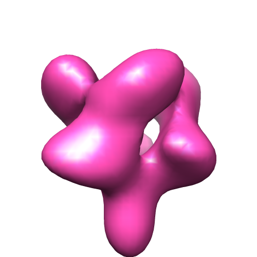 EMD-2065.png EMD-2065.png | 734.4 KB | ||
| Archive directory |  http://ftp.pdbj.org/pub/emdb/structures/EMD-2065ftp://ftp.pdbj.org/pub/emdb/structures/EMD-2065 http://ftp.pdbj.org/pub/emdb/structures/EMD-2065ftp://ftp.pdbj.org/pub/emdb/structures/EMD-2065 | HTTPS FTP |
-Related structure data


-Links
| EMDB pages | EMDB (EBI/PDBe) / EMDataResource |
|---|
-Map
| File | Download / File: emd_2065.map.gz / Format: CCP4 / Size: 422.9 KB / Type: IMAGE STORED AS FLOATING POINT NUMBER (4 BYTES) | ||||||||||||||||||||||||||||||||||||||||||||||||||||||||||||||||||||
|---|---|---|---|---|---|---|---|---|---|---|---|---|---|---|---|---|---|---|---|---|---|---|---|---|---|---|---|---|---|---|---|---|---|---|---|---|---|---|---|---|---|---|---|---|---|---|---|---|---|---|---|---|---|---|---|---|---|---|---|---|---|---|---|---|---|---|---|---|---|
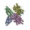
| Annotation | Reconstruction of the R-peptide precursor of Moloney murine leukemia virus Env in its isomerization arrested intermediate state | ||||||||||||||||||||||||||||||||||||||||||||||||||||||||||||||||||||
| Projections & slices | Image control
Images are generated by Spider. | ||||||||||||||||||||||||||||||||||||||||||||||||||||||||||||||||||||
| Voxel size | X=Y=Z: 3.5 Å | ||||||||||||||||||||||||||||||||||||||||||||||||||||||||||||||||||||
| Density |
| ||||||||||||||||||||||||||||||||||||||||||||||||||||||||||||||||||||
| Symmetry | Space group: 1 | ||||||||||||||||||||||||||||||||||||||||||||||||||||||||||||||||||||
| Details | EMDB XML:
CCP4 map header:
| ||||||||||||||||||||||||||||||||||||||||||||||||||||||||||||||||||||
 Z (Sec.)
 Y (Row.)
 X (Col.)


-Supplemental data
- Sample components
-Entire : Isomerization arrested intermediate state of the R-peptide precur...
| Entire | Name: Isomerization arrested intermediate state of the R-peptide precursor of Moloney murine leukemia virus Env |
|---|---|
| Components |
|
-Supramolecule #1000: Isomerization arrested intermediate state of the R-peptide precur...
| Supramolecule | Name: Isomerization arrested intermediate state of the R-peptide precursor of Moloney murine leukemia virus Env type: sample / ID: 1000 / Oligomeric state: trimeric / Number unique components: 1 |
|---|---|
| Molecular weight | Experimental: 500 KDa / Theoretical: 270 KDa / Method: Blue native PAGE |
-Macromolecule #1: (gp70-Pr15E)3
| Macromolecule | Name: (gp70-Pr15E)3 / type: protein_or_peptide / ID: 1 / Name.synonym: (SU-TM)3 ; Env Details: Expression system cell line Human Embryonic Kidney 293T Number of copies: 3 / Oligomeric state: trimeric / Recombinant expression: Yes |
|---|---|
| Source (natural) | Organism: Moloney murine leukemia virus / synonym: Mo-MLV |
| Molecular weight | Experimental: 500 KDa / Theoretical: 270 KDa |
| Recombinant expression | Organism:  Homo sapiens (human) / Recombinant plasmid: pNCA Homo sapiens (human) / Recombinant plasmid: pNCA |
-Experimental details
-Structure determination
| Method | negative staining, cryo EM |
|---|---|
 Processing Processing | single particle reconstruction |
| Aggregation state | particle |
-Sample preparation
| Concentration | 0.1 mg/mL |
|---|---|
| Buffer | pH: 7.4 Details: 50 mM Hepes, 100 mM NaCl, 1,8 mM CaCl2, 0.05% Triton X-100 |
| Staining | Type: NEGATIVE Details: The specimen was frozen in liquid ethane and transferred to liquid nitrogen for EM inspection without staining. |
| Grid | Details: 400 mesh holey carbon grid. The grids were glow discharged. |
| Vitrification | Cryogen name: ETHANE / Chamber humidity: 99 % / Chamber temperature: 77 K / Instrument: FEI VITROBOT MARK II Timed resolved state: Vitrified 45 msec after spraying with effector Method: Blot for 3 seconds before plunging |
- Electron microscopy
| Microscope | JEOL 2100F |
|---|---|
| Temperature | Min: 93 K / Max: 96 K / Average: 95 K |
| Alignment procedure | Legacy - Astigmatism: Objective lens astigmatism was corrected using online FFT |
| Date | Jan 21, 2011 |
| Image recording | Category: CCD / Film or detector model: GENERIC CCD / Digitization - Sampling interval: 3.5 µm / Number real images: 687 / Average electron dose: 9 e/Å2 / Details: The images were recorded by CCD camera / Bits/pixel: 14 |
| Tilt angle min | 0 |
| Tilt angle max | 0 |
| Electron beam | Acceleration voltage: 200 kV / Electron source:  FIELD EMISSION GUN FIELD EMISSION GUN |
| Electron optics | Calibrated magnification: 43200 / Illumination mode: SPOT SCAN / Imaging mode: BRIGHT FIELD / Cs: 2.0 mm / Nominal defocus max: 4.0 µm / Nominal defocus min: 2.5 µm / Nominal magnification: 43200 |
| Sample stage | Specimen holder: Liquid nitrogen cooled / Specimen holder model: GATAN LIQUID NITROGEN |
-Image processing
| Details | The particles were selected using an automatic selection program and the damaged particles were removed by visual inspection |
|---|---|
| CTF correction | Details: Each particle |
| Final reconstruction | Applied symmetry - Point group: C3 (3 fold cyclic) / Algorithm: OTHER / Resolution.type: BY AUTHOR / Resolution: 22.0 Å / Resolution method: FSC 0.5 CUT-OFF / Software - Name: EMAN1, EMAN2 Details: Final maps were calculated from seven averaged datasets Number images used: 3601 |
| Final two d classification | Number classes: 131 |
-Atomic model buiding 1
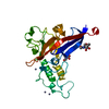
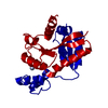
| Initial model | PDB ID: Chain - Chain ID: A |
|---|---|
| Software | Name: Chimera, O |
| Details | Protocol: Rigid body. The atomic model for the Mo-RBD was obtained using the atomic structure of the highly homologous F-RBD (Fass et al, 1997) (Protein Data Bank ID, 1AOL) and the SWISS-MODEL protein structure homology-modeling server (accessible through the ExPASy web server). |
| Refinement | Space: REAL / Protocol: RIGID BODY FIT |