 Movie
Movie Controller

[English] 日本語
 Yorodumi
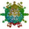
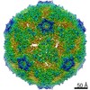
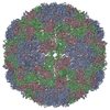
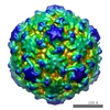
- EMDB-20474: Poliovirus Type-1 Mahoney receptor catalysed 135S particle incuba... -
+ Open data

- Basic information
| Entry | Database: EMDB / ID: EMD-20474 | |||||||||
|---|---|---|---|---|---|---|---|---|---|---|
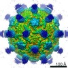
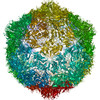
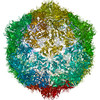
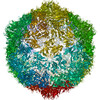
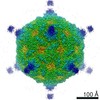
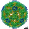
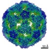
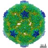
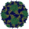
| Title | Poliovirus Type-1 Mahoney receptor catalysed 135S particle incubated with anti-VP1 mAb for 1 hour at 37 degrees C | |||||||||
 Map data Map data | Poliovirus Type-1 Mahoney receptor catalysed 135S particle incubated with anti-VP1 mAb for 1 hr at 37% u02DAC. | |||||||||
 Sample Sample |
| |||||||||
| Biological species |   Poliovirus type 1 (strain Mahoney) Poliovirus type 1 (strain Mahoney) | |||||||||
| Method | single particle reconstruction / cryo EM / Resolution: 3.6 Å | |||||||||
 Authors Authors | Hogle JM / Filman DJ / Shah PNM | |||||||||
| Funding support |  United States, 1 items United States, 1 items
| |||||||||
 Citation Citation | Journal: PLoS Pathog / Year: 2020 Title: Cryo-EM structures reveal two distinct conformational states in a picornavirus cell entry intermediate. Authors: Pranav N M Shah / David J Filman / Krishanthi S Karunatilaka / Emma L Hesketh / Elisabetta Groppelli / Mike Strauss / James M Hogle /   Abstract: The virions of enteroviruses such as poliovirus undergo a global conformational change after binding to the cellular receptor, characterized by a 4% expansion, and by the opening of holes at the two ...The virions of enteroviruses such as poliovirus undergo a global conformational change after binding to the cellular receptor, characterized by a 4% expansion, and by the opening of holes at the two and quasi-three-fold symmetry axes of the capsid. The resultant particle is called a 135S particle or A-particle and is thought to be on the pathway to a productive infection. Previously published studies have concluded that the membrane-interactive peptides, namely VP4 and the N-terminus of VP1, are irreversibly externalized in the 135S particle. However, using established protocols to produce the 135S particle, and single particle cryo-electron microscopy methods, we have identified at least two unique states that we call the early and late 135S particle. Surprisingly, only in the "late" 135S particles have detectable levels of the VP1 N-terminus been trapped outside the capsid. Moreover, we observe a distinct density inside the capsid that can be accounted for by VP4 that remains associated with the genome. Taken together our results conclusively demonstrate that the 135S particle is not a unique conformation, but rather a family of conformations that could exist simultaneously. | |||||||||
| History |
|
- Structure visualization
| Movie |
 Movie viewer Movie viewer |
|---|---|
| Structure viewer | EM map: SurfViewMolmilJmol/JSmol |
| Supplemental images |

- Downloads & links
-EMDB archive
| Map data | emd_20474.map.gz | 253.8 MB | EMDB map data format | |
|---|---|---|---|---|
| Header (meta data) | emd-20474-v30.xmlemd-20474.xml | 11.4 KB 11.4 KB | Display Display | EMDB header |
| FSC (resolution estimation) | emd_20474_fsc.xml | 15.9 KB | Display | FSC data file |
| Images | 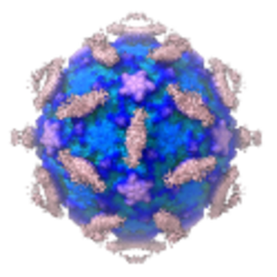 emd_20474.png emd_20474.png | 178.3 KB | ||
| Archive directory |  http://ftp.pdbj.org/pub/emdb/structures/EMD-20474ftp://ftp.pdbj.org/pub/emdb/structures/EMD-20474 http://ftp.pdbj.org/pub/emdb/structures/EMD-20474ftp://ftp.pdbj.org/pub/emdb/structures/EMD-20474 | HTTPS FTP |
-Related structure data

-Links
| EMDB pages | EMDB (EBI/PDBe) / EMDataResource |
|---|
-Map
| File | Download / File: emd_20474.map.gz / Format: CCP4 / Size: 343 MB / Type: IMAGE STORED AS FLOATING POINT NUMBER (4 BYTES) | ||||||||||||||||||||||||||||||||||||||||||||||||||||||||||||||||||||
|---|---|---|---|---|---|---|---|---|---|---|---|---|---|---|---|---|---|---|---|---|---|---|---|---|---|---|---|---|---|---|---|---|---|---|---|---|---|---|---|---|---|---|---|---|---|---|---|---|---|---|---|---|---|---|---|---|---|---|---|---|---|---|---|---|---|---|---|---|---|
| Annotation | Poliovirus Type-1 Mahoney receptor catalysed 135S particle incubated with anti-VP1 mAb for 1 hr at 37% u02DAC. | ||||||||||||||||||||||||||||||||||||||||||||||||||||||||||||||||||||
| Projections & slices | Image control
Images are generated by Spider. | ||||||||||||||||||||||||||||||||||||||||||||||||||||||||||||||||||||
| Voxel size | X=Y=Z: 1.11 Å | ||||||||||||||||||||||||||||||||||||||||||||||||||||||||||||||||||||
| Density |
| ||||||||||||||||||||||||||||||||||||||||||||||||||||||||||||||||||||
| Symmetry | Space group: 1 | ||||||||||||||||||||||||||||||||||||||||||||||||||||||||||||||||||||
| Details | EMDB XML:
CCP4 map header:
| ||||||||||||||||||||||||||||||||||||||||||||||||||||||||||||||||||||
 Z (Sec.)
 Y (Row.)
 X (Col.)


-Supplemental data
- Sample components
-Entire : Poliovirus type 1 (strain Mahoney)
| Entire | Name: Poliovirus type 1 (strain Mahoney) |
|---|---|
| Components |
|
-Supramolecule #1: Poliovirus type 1 (strain Mahoney)
| Supramolecule | Name: Poliovirus type 1 (strain Mahoney) / type: virus / ID: 1 / Parent: 0 / Macromolecule list: #1-#3 / NCBI-ID: 12081 / Sci species name: Poliovirus type 1 (strain Mahoney) / Virus type: VIRION / Virus isolate: STRAIN / Virus enveloped: No / Virus empty: No |
|---|---|
| Host (natural) | Organism:  Homo sapiens (human) Homo sapiens (human) |
| Molecular weight | Theoretical: 10.0 MDa |
| Virus shell | Shell ID: 1 / Name: Capsid / Diameter: 300.0 Å / T number (triangulation number): 1 |
-Experimental details
-Structure determination
| Method | cryo EM |
|---|---|
 Processing Processing | single particle reconstruction |
| Aggregation state | particle |
-Sample preparation
| Concentration | 0.4 mg/mL |
|---|---|
| Buffer | pH: 7.5 / Details: 20 mM Tris-HCl, pH 7.5 + 2 mM CaCl2 |
| Grid | Support film - Material: CARBON / Support film - topology: LACEY / Details: unspecified |
| Vitrification | Cryogen name: ETHANE / Chamber humidity: 100 % / Chamber temperature: 277 K / Instrument: FEI VITROBOT MARK IV |
| Details | This sample was monodisperse. |
- Electron microscopy
| Microscope | FEI TECNAI ARCTICA |
|---|---|
| Image recording | Film or detector model: GATAN K3 (6k x 4k) / Detector mode: COUNTING / Number grids imaged: 1 / Average electron dose: 1.06 e/Å2 |
| Electron beam | Acceleration voltage: 200 kV / Electron source:  FIELD EMISSION GUN FIELD EMISSION GUN |
| Electron optics | Illumination mode: SPOT SCAN / Imaging mode: BRIGHT FIELD |
| Sample stage | Specimen holder model: FEI TITAN KRIOS AUTOGRID HOLDER / Cooling holder cryogen: NITROGEN |
| Experimental equipment |  Model: Talos Arctica / Image courtesy: FEI Company |
+Image processing

-Atomic model buiding 1
| Refinement | Space: REAL / Protocol: FLEXIBLE FIT |
|---|
