 Movie
Movie Controller

[English] 日本語
 Yorodumi
- EMDB-2036: Three-dimensional structure of Tripeptidyl peptidase II from Homo... -
+ Open data

- Basic information
| Entry | Database: EMDB / ID: EMD-2036 | |||||||||
|---|---|---|---|---|---|---|---|---|---|---|
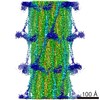
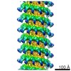
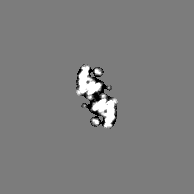
| Title | Three-dimensional structure of Tripeptidyl peptidase II from Homo sapiens - a spindle-shaped homo-36mer | |||||||||
 Map data Map data | Tripeptidyl peptidase II complex of Homo sapiens | |||||||||
 Sample Sample |
| |||||||||
| Function / homology | Peptidase S8/S53 domain Function and homology information Function and homology information | |||||||||
| Biological species |  Homo sapiens (human) Homo sapiens (human) | |||||||||
| Method | single particle reconstruction / cryo EM / Resolution: 9.9 Å | |||||||||
 Authors Authors | Schoenegge AM / Villa E / Foerster F / Hegerl R / Peters J / Baumeister W / Rockel B | |||||||||
 Citation Citation | Journal: Structure / Year: 2012 Title: The structure of human tripeptidyl peptidase II as determined by a hybrid approach. Authors: Anne-Marie Schönegge / Elizabeth Villa / Friedrich Förster / Reiner Hegerl / Jürgen Peters / Wolfgang Baumeister / Beate Rockel /  Abstract: Tripeptidyl-peptidase II (TPPII) is a high molecular mass (∼5 MDa) serine protease, which is thought to act downstream of the 26S proteasome, cleaving peptides released by the latter. Here, the ...Tripeptidyl-peptidase II (TPPII) is a high molecular mass (∼5 MDa) serine protease, which is thought to act downstream of the 26S proteasome, cleaving peptides released by the latter. Here, the structure of human TPPII (HsTPPII) has been determined to subnanometer resolution by cryoelectron microscopy and single-particle analysis. The complex is built from two strands forming a quasihelical structure harboring a complex system of inner cavities. HsTPPII particles exhibit some polymorphism resulting in complexes consisting of nine or of eight dimers per strand. To obtain deeper insights into the architecture and function of HsTPPII, we have created a pseudoatomic structure of the HsTPPII spindle using a comparative model of HsTPPII dimers and molecular dynamics flexible fitting. Analyses of the resulting hybrid structure of the HsTPPII holocomplex provide new insights into the mechanism of maturation and activation. | |||||||||
| History |
|
- Structure visualization
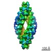
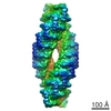
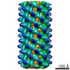
| Movie |
Movie viewer |
|---|---|
| Structure viewer | EM map: SurfViewMolmilJmol/JSmol |
| Supplemental images |

- Downloads & links
-EMDB archive
| Map data | emd_2036.map.gz | 22.1 MB | EMDB map data format | |
|---|---|---|---|---|
| Header (meta data) | emd-2036-v30.xmlemd-2036.xml | 8.4 KB 8.4 KB | Display Display | EMDB header |
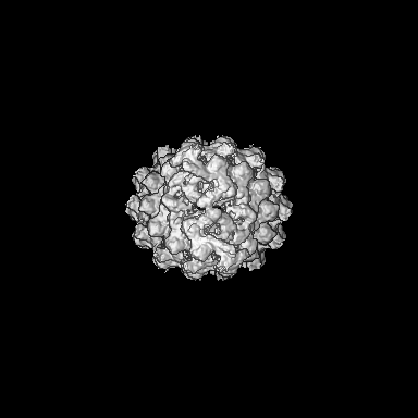
| Images | 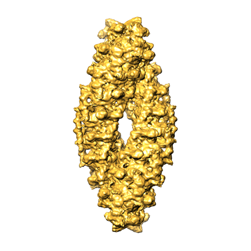 EMD_2036.png EMD_2036.png | 150.2 KB | ||
| Archive directory |  http://ftp.pdbj.org/pub/emdb/structures/EMD-2036ftp://ftp.pdbj.org/pub/emdb/structures/EMD-2036 http://ftp.pdbj.org/pub/emdb/structures/EMD-2036ftp://ftp.pdbj.org/pub/emdb/structures/EMD-2036 | HTTPS FTP |
-Validation report
| Summary document | emd_2036_validation.pdf.gz | 217.9 KB | Display | EMDB validaton report |
|---|---|---|---|---|
| Full document | emd_2036_full_validation.pdf.gz | 217 KB | Display | |
| Data in XML | emd_2036_validation.xml.gz | 7.1 KB | Display | |
| Arichive directory | https://ftp.pdbj.org/pub/emdb/validation_reports/EMD-2036ftp://ftp.pdbj.org/pub/emdb/validation_reports/EMD-2036 | HTTPS FTP |
-Related structure data


-Links
| EMDB pages | EMDB (EBI/PDBe) / EMDataResource |
|---|
-Map
| File | Download / File: emd_2036.map.gz / Format: CCP4 / Size: 210.9 MB / Type: IMAGE STORED AS FLOATING POINT NUMBER (4 BYTES) | ||||||||||||||||||||||||||||||||||||||||||||||||||||||||||||||||||||
|---|---|---|---|---|---|---|---|---|---|---|---|---|---|---|---|---|---|---|---|---|---|---|---|---|---|---|---|---|---|---|---|---|---|---|---|---|---|---|---|---|---|---|---|---|---|---|---|---|---|---|---|---|---|---|---|---|---|---|---|---|---|---|---|---|---|---|---|---|---|
| Annotation | Tripeptidyl peptidase II complex of Homo sapiens | ||||||||||||||||||||||||||||||||||||||||||||||||||||||||||||||||||||
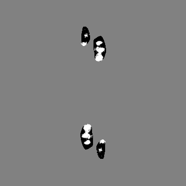
| Projections & slices | Image control
Images are generated by Spider. | ||||||||||||||||||||||||||||||||||||||||||||||||||||||||||||||||||||
| Voxel size | X=Y=Z: 1.78 Å | ||||||||||||||||||||||||||||||||||||||||||||||||||||||||||||||||||||
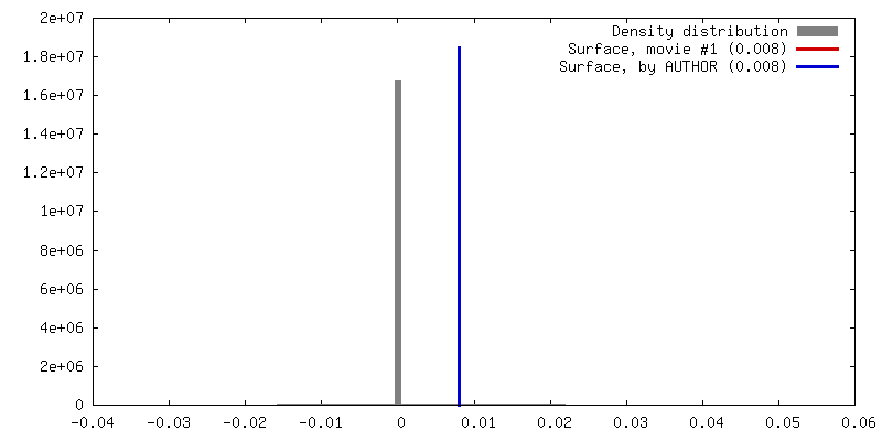
| Density |
| ||||||||||||||||||||||||||||||||||||||||||||||||||||||||||||||||||||
| Symmetry | Space group: 1 | ||||||||||||||||||||||||||||||||||||||||||||||||||||||||||||||||||||
| Details | EMDB XML:
CCP4 map header:
| ||||||||||||||||||||||||||||||||||||||||||||||||||||||||||||||||||||
 Z (Sec.)
 Y (Row.)
 X (Col.)


-Supplemental data
- Sample components
-Entire : Tripeptidyl peptidase II of Homo Sapiens
| Entire | Name: Tripeptidyl peptidase II of Homo Sapiens |
|---|---|
| Components |
|
-Supramolecule #1000: Tripeptidyl peptidase II of Homo Sapiens
| Supramolecule | Name: Tripeptidyl peptidase II of Homo Sapiens / type: sample / ID: 1000 / Oligomeric state: 36-mer / Number unique components: 1 |
|---|---|
| Molecular weight | Theoretical: 5 MDa |
-Macromolecule #1: Tripeptidyl peptidase II
| Macromolecule | Name: Tripeptidyl peptidase II / type: protein_or_peptide / ID: 1 / Name.synonym: Tripeptidyl peptidase II of Homo Sapiens / Number of copies: 1 / Oligomeric state: 36-mer / Recombinant expression: Yes |
|---|---|
| Source (natural) | Organism: Homo sapiens (human) / synonym: Human |
| Molecular weight | Theoretical: 5 MDa |
| Recombinant expression | Organism:  |
| Sequence | InterPro: Peptidase S8/S53 domain |
-Experimental details
-Structure determination
| Method | cryo EM |
|---|---|
 Processing Processing | single particle reconstruction |
| Aggregation state | particle |
-Sample preparation
| Concentration | 0.2 mg/mL |
|---|---|
| Buffer | pH: 7.5 / Details: 80 mM KPO4, 1 mM DTT, 5% glycerol |
| Grid | Details: glow-discharged C-flat 4/1 grids covered with a thin carbon film |
| Vitrification | Cryogen name: ETHANE / Instrument: OTHER Method: Sample was applied on grid, blotted briefly and washed twice with buffer (40 mM ammonium sulfate, pH 7.5) |
- Electron microscopy
| Microscope | FEI TECNAI F20 |
|---|---|
| Image recording | Category: CCD / Film or detector model: FEI EAGLE (2k x 2k) / Average electron dose: 15 e/Å2 |
| Electron beam | Acceleration voltage: 200 kV / Electron source:  FIELD EMISSION GUN FIELD EMISSION GUN |
| Electron optics | Calibrated magnification: 84270 / Illumination mode: FLOOD BEAM / Imaging mode: BRIGHT FIELD / Cs: 2 mm |
| Sample stage | Specimen holder: side-entry cryoholder / Specimen holder model: GATAN LIQUID NITROGEN |
| Experimental equipment |  Model: Tecnai F20 / Image courtesy: FEI Company |
-Image processing
| Details | Particles were selected manually |
|---|---|
| CTF correction | Details: each image |
| Final reconstruction | Applied symmetry - Point group: D2 (2x2 fold dihedral) / Algorithm: OTHER / Resolution.type: BY AUTHOR / Resolution: 9.9 Å / Resolution method: FSC 0.5 CUT-OFF / Software - Name: XMIPP / Details: Final map was b-factor corrected. / Number images used: 96116 |
