 Movie
Movie Controller


+ Open data

- Basic information
| Entry | Database: EMDB / ID: EMD-4119 | |||||||||
|---|---|---|---|---|---|---|---|---|---|---|
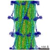
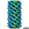
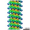
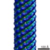
| Title | In situ TPPII 36mer | |||||||||
 Map data Map data | TPPII 36mer | |||||||||
 Sample Sample |
| |||||||||
| Biological species |  | |||||||||
| Method | subtomogram averaging / cryo EM / Resolution: 27.8 Å | |||||||||
 Authors Authors | Fukuda Y / Beck F / Baumeister W | |||||||||
 Citation Citation | Journal: Proc Natl Acad Sci U S A / Year: 2017 Title: In situ structural studies of tripeptidyl peptidase II (TPPII) reveal spatial association with proteasomes. Authors: Yoshiyuki Fukuda / Florian Beck / Jürgen M Plitzko / Wolfgang Baumeister /  Abstract: Tripeptidyl peptidase II (TPPII) is a eukaryotic protease acting downstream of the 26S proteasome; it removes tripeptides from the degradation products released by the proteasome. Structural studies ...Tripeptidyl peptidase II (TPPII) is a eukaryotic protease acting downstream of the 26S proteasome; it removes tripeptides from the degradation products released by the proteasome. Structural studies in vitro have revealed the basic architecture of TPPII, a two-stranded linear polymer that assembles to form a spindle-shaped complex of ∼6 MDa. Dependent on protein concentration, TPPII has a distinct tendency for polymorphism. Therefore, its structure in vivo has remained unclear. To resolve this issue, we have scrutinized cryo-electron tomograms of rat hippocampal neurons for the occurrence and spatial distribution of TPPII by template matching. The quality of the tomograms recorded with the Volta phase plate enabled a detailed structural analysis of TPPII despite its low abundance. Two different assembly states (36-mers and 32-mers) coexist as well as occasional extended forms with longer strands. A distance analysis of the relative locations of TPPII and 26S proteasomes confirmed the visual impression that these two complexes spatially associate in agreement with TPPII's role in postproteasomal degradation. | |||||||||
| History |
|
- Structure visualization
| Movie |
 Movie viewer Movie viewer |
|---|---|
| Structure viewer | EM map: SurfViewMolmilJmol/JSmol |
| Supplemental images |


- Downloads & links
-EMDB archive
| Map data | emd_4119.map.gz | 27.6 MB | EMDB map data format | |
|---|---|---|---|---|
| Header (meta data) | emd-4119-v30.xmlemd-4119.xml | 11.3 KB 11.3 KB | Display Display | EMDB header |
| Images | 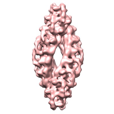 emd_4119.jpg emd_4119.jpg emd_4119.png emd_4119.png | 24 KB 131 KB | ||
| Archive directory |  http://ftp.pdbj.org/pub/emdb/structures/EMD-4119ftp://ftp.pdbj.org/pub/emdb/structures/EMD-4119 http://ftp.pdbj.org/pub/emdb/structures/EMD-4119ftp://ftp.pdbj.org/pub/emdb/structures/EMD-4119 | HTTPS FTP |
-Related structure data
-Links
| EMDB pages | EMDB (EBI/PDBe) / EMDataResource |
|---|
-Map
| File | Download / File: emd_4119.map.gz / Format: CCP4 / Size: 30.5 MB / Type: IMAGE STORED AS FLOATING POINT NUMBER (4 BYTES) | ||||||||||||||||||||||||||||||||||||||||||||||||||||||||||||||||||||
|---|---|---|---|---|---|---|---|---|---|---|---|---|---|---|---|---|---|---|---|---|---|---|---|---|---|---|---|---|---|---|---|---|---|---|---|---|---|---|---|---|---|---|---|---|---|---|---|---|---|---|---|---|---|---|---|---|---|---|---|---|---|---|---|---|---|---|---|---|---|
| Annotation | TPPII 36mer | ||||||||||||||||||||||||||||||||||||||||||||||||||||||||||||||||||||
| Projections & slices | Image control
Images are generated by Spider. | ||||||||||||||||||||||||||||||||||||||||||||||||||||||||||||||||||||
| Voxel size | X=Y=Z: 4.21 Å | ||||||||||||||||||||||||||||||||||||||||||||||||||||||||||||||||||||
| Density |
| ||||||||||||||||||||||||||||||||||||||||||||||||||||||||||||||||||||
| Symmetry | Space group: 1 | ||||||||||||||||||||||||||||||||||||||||||||||||||||||||||||||||||||
| Details | EMDB XML:
CCP4 map header:
| ||||||||||||||||||||||||||||||||||||||||||||||||||||||||||||||||||||
 Z (Sec.)
 Y (Row.)
 X (Col.)


-Supplemental data
- Sample components
-Entire : Tripeptidyl peptidase II
| Entire | Name: Tripeptidyl peptidase II |
|---|---|
| Components |
|
-Supramolecule #1: Tripeptidyl peptidase II
| Supramolecule | Name: Tripeptidyl peptidase II / type: complex / ID: 1 / Parent: 0 / Details: 36mer |
|---|---|
| Source (natural) | Organism: |
| Molecular weight | Theoretical: 5 MDa |
-Experimental details
-Structure determination
| Method | cryo EM |
|---|---|
 Processing Processing | subtomogram averaging |
| Aggregation state | cell |
-Sample preparation
| Buffer | pH: 7 |
|---|---|
| Grid | Model: Quantifoil R1/4 / Material: GOLD / Mesh: 200 / Support film - Material: CARBON / Support film - topology: HOLEY ARRAY / Support film - Film thickness: 1000.0 nm |
| Vitrification | Cryogen name: ETHANE-PROPANE / Chamber humidity: 90 % / Chamber temperature: 310 K / Instrument: FEI VITROBOT MARK IV |
| Details | in situ |
- Electron microscopy
| Microscope | FEI TITAN KRIOS |
|---|---|
| Temperature | Min: 70.0 K / Max: 70.0 K |
| Specialist optics | Phase plate: VOLTA PHASE PLATE / Energy filter - Name: GIF / Energy filter - Lower energy threshold: 0 eV / Energy filter - Upper energy threshold: 20 eV |
| Image recording | Film or detector model: GATAN K2 SUMMIT (4k x 4k) / Detector mode: COUNTING / Digitization - Dimensions - Width: 3838 pixel / Digitization - Dimensions - Height: 3710 pixel / Digitization - Sampling interval: 5.0 µm / Digitization - Frames/image: 1-10 / Number grids imaged: 2 / Average exposure time: 1.4 sec. / Average electron dose: 1.8 e/Å2 |
| Electron beam | Acceleration voltage: 300 kV / Electron source:  FIELD EMISSION GUN FIELD EMISSION GUN |
| Electron optics | C2 aperture diameter: 70.0 µm / Calibrated defocus max: 1.0 µm / Calibrated defocus min: 0.5 µm / Illumination mode: FLOOD BEAM / Imaging mode: BRIGHT FIELD / Cs: 2.7 mm / Nominal defocus max: 1.0 µm / Nominal defocus min: 0.5 µm / Nominal magnification: 33000 |
| Sample stage | Specimen holder model: FEI TITAN KRIOS AUTOGRID HOLDER / Cooling holder cryogen: NITROGEN |
| Experimental equipment |  Model: Titan Krios / Image courtesy: FEI Company |
-Image processing
| Final reconstruction | Applied symmetry - Point group: D2 (2x2 fold dihedral) / Resolution.type: BY AUTHOR / Resolution: 27.8 Å / Resolution method: FSC 0.143 CUT-OFF / Software - Name: PyTom (ver. 0.97) / Number subtomograms used: 81 |
|---|---|
| Extraction | Number tomograms: 70 / Number images used: 81 / Reference model: emdb 2036 / Method: template matching / Software - Name: PyTom (ver. 0.97) |
| Final angle assignment | Type: OTHER / Software - Name: PyTom (ver. 0.97) / Details: subtomogram averaging |
