 Movie
Movie Controller

[English] 日本語
 Yorodumi
- EMDB-1732: Three-dimensional structure of Tripeptidyl peptidase II from Dros... -
+ Open data

- Basic information
| Entry | Database: EMDB / ID: EMD-1732 | |||||||||
|---|---|---|---|---|---|---|---|---|---|---|
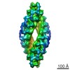
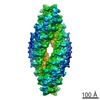
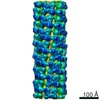
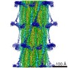
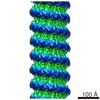
| Title | Three-dimensional structure of Tripeptidyl peptidase II from Drosophila melanogaster - a spindle shaped homo-40mer | |||||||||
 Map data Map data | EM density map of TPPII from Drosophila melanogaster | |||||||||
 Sample Sample |
| |||||||||
 Keywords Keywords | Protease / subtilase | |||||||||
| Biological species |  | |||||||||
| Method | single particle reconstruction / cryo EM / Resolution: 14.0 Å | |||||||||
 Authors Authors | Chuang CK / Rockel B / Seyit G / Walian PJ / Schoenegge A-M / Peters J / Zwart PH / Baumeister W / Jap BK | |||||||||
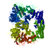
 Citation Citation | Journal: Nat Struct Mol Biol / Year: 2010 Title: Hybrid molecular structure of the giant protease tripeptidyl peptidase II. Authors: Crystal K Chuang / Beate Rockel / Gönül Seyit / Peter J Walian / Anne-Marie Schönegge / Jürgen Peters / Petrus H Zwart / Wolfgang Baumeister / Bing K Jap /  Abstract: Tripeptidyl peptidase II (TPP II) is the largest known eukaryotic protease (6 MDa). It is believed to act downstream of the 26S proteasome, cleaving tripeptides from the N termini of longer peptides, ...Tripeptidyl peptidase II (TPP II) is the largest known eukaryotic protease (6 MDa). It is believed to act downstream of the 26S proteasome, cleaving tripeptides from the N termini of longer peptides, and it is implicated in numerous cellular processes. Here we report the structure of Drosophila TPP II determined by a hybrid approach. We solved the structure of the dimer by X-ray crystallography and docked it into the three-dimensional map of the holocomplex, which we obtained by single-particle cryo-electron microscopy. The resulting structure reveals the compartmentalization of the active sites inside a system of chambers and suggests the existence of a molecular ruler determining the size of the cleavage products. Furthermore, the structure suggests a model for activation of TPP II involving the relocation of a flexible loop and a repositioning of the active-site serine, coupling it to holocomplex assembly and active-site sequestration. | |||||||||
| History |
|
- Structure visualization
| Movie |
 Movie viewer Movie viewer |
|---|---|
| Structure viewer | EM map: SurfViewMolmilJmol/JSmol |
| Supplemental images |

- Downloads & links
-EMDB archive
| Map data | emd_1732.map.gz | 113.3 MB | EMDB map data format | |
|---|---|---|---|---|
| Header (meta data) | emd-1732-v30.xmlemd-1732.xml | 8.4 KB 8.4 KB | Display Display | EMDB header |
| Images | 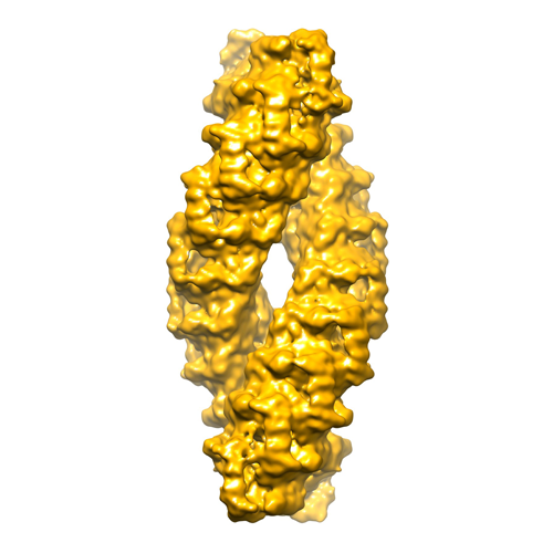 emd1732.png emd1732.png | 136.8 KB | ||
| Archive directory |  http://ftp.pdbj.org/pub/emdb/structures/EMD-1732ftp://ftp.pdbj.org/pub/emdb/structures/EMD-1732 http://ftp.pdbj.org/pub/emdb/structures/EMD-1732ftp://ftp.pdbj.org/pub/emdb/structures/EMD-1732 | HTTPS FTP |
-Related structure data
-Links
| EMDB pages | EMDB (EBI/PDBe) / EMDataResource |
|---|
-Map
| File | Download / File: emd_1732.map.gz / Format: CCP4 / Size: 122.1 MB / Type: IMAGE STORED AS FLOATING POINT NUMBER (4 BYTES) | ||||||||||||||||||||||||||||||||||||||||||||||||||||||||||||||||||||
|---|---|---|---|---|---|---|---|---|---|---|---|---|---|---|---|---|---|---|---|---|---|---|---|---|---|---|---|---|---|---|---|---|---|---|---|---|---|---|---|---|---|---|---|---|---|---|---|---|---|---|---|---|---|---|---|---|---|---|---|---|---|---|---|---|---|---|---|---|---|
| Annotation | EM density map of TPPII from Drosophila melanogaster | ||||||||||||||||||||||||||||||||||||||||||||||||||||||||||||||||||||
| Projections & slices | Image control
Images are generated by Spider. | ||||||||||||||||||||||||||||||||||||||||||||||||||||||||||||||||||||
| Voxel size | X=Y=Z: 2.16 Å | ||||||||||||||||||||||||||||||||||||||||||||||||||||||||||||||||||||
| Density |
| ||||||||||||||||||||||||||||||||||||||||||||||||||||||||||||||||||||
| Symmetry | Space group: 1 | ||||||||||||||||||||||||||||||||||||||||||||||||||||||||||||||||||||
| Details | EMDB XML:
CCP4 map header:
| ||||||||||||||||||||||||||||||||||||||||||||||||||||||||||||||||||||
 Z (Sec.)
 Y (Row.)
 X (Col.)


-Supplemental data
- Sample components
-Entire : Tripeptidyl peptidase II complex of Drosophila melanogaster
| Entire | Name: Tripeptidyl peptidase II complex of Drosophila melanogaster |
|---|---|
| Components |
|
-Supramolecule #1000: Tripeptidyl peptidase II complex of Drosophila melanogaster
| Supramolecule | Name: Tripeptidyl peptidase II complex of Drosophila melanogaster type: sample / ID: 1000 / Oligomeric state: 40-mer / Number unique components: 1 |
|---|---|
| Molecular weight | Theoretical: 6 MDa |
-Macromolecule #1: Tripeptidyl peptidase II
| Macromolecule | Name: Tripeptidyl peptidase II / type: protein_or_peptide / ID: 1 / Name.synonym: TPPII / Number of copies: 40 / Oligomeric state: 40-mer / Recombinant expression: Yes |
|---|---|
| Source (natural) | Organism: |
| Molecular weight | Theoretical: 150 KDa |
| Recombinant expression | Organism:  |
-Experimental details
-Structure determination
| Method | cryo EM |
|---|---|
 Processing Processing | single particle reconstruction |
| Aggregation state | particle |
-Sample preparation
| Buffer | pH: 7.8 / Details: 40 mM KPO4, pH 7.8, 2 mM DTT, 5% glycerol |
|---|---|
| Grid | Details: Holey carbon 200 mesh copper grids covered with thin carbon film |
| Vitrification | Cryogen name: ETHANE / Instrument: OTHER Method: Sample was applied on grid, blotted briefly and washed twice with buffer (40 mM ammonium sulfate, pH 7.5) |
- Electron microscopy
| Microscope | FEI/PHILIPS CM200FEG |
|---|---|
| Image recording | Category: CCD / Film or detector model: GENERIC TVIPS (4k x 4k) / Average electron dose: 15 e/Å2 |
| Electron beam | Acceleration voltage: 160 kV / Electron source:  FIELD EMISSION GUN FIELD EMISSION GUN |
| Electron optics | Calibrated magnification: 69500 / Illumination mode: FLOOD BEAM / Imaging mode: BRIGHT FIELD / Nominal defocus max: 3.0 µm / Nominal defocus min: 0.7 µm |
| Sample stage | Specimen holder: Side-entry / Specimen holder model: GATAN LIQUID NITROGEN |
-Image processing
| CTF correction | Details: Each CCD frame |
|---|---|
| Final reconstruction | Applied symmetry - Point group: D2 (2x2 fold dihedral) / Resolution.type: BY AUTHOR / Resolution: 14.0 Å / Resolution method: FSC 0.5 CUT-OFF / Software - Name: EMAN / Number images used: 37195 |
