 Movie
Movie Controller


+ Open data

- Basic information
| Entry | Database: EMDB / ID: EMD-20223 | ||||||||||||
|---|---|---|---|---|---|---|---|---|---|---|---|---|---|
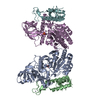
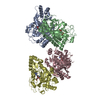
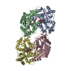
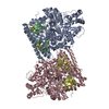
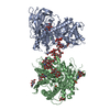
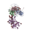
| Title | Structure of the Rhodopsin-Transducin-Nanobody Complex | ||||||||||||
 Map data Map data | Rhodopsin-Transducin-Nanobody Complex | ||||||||||||
 Sample Sample |
| ||||||||||||
 Keywords Keywords | GPCR / G protein / Complex / SIGNALING PROTEIN-IMMUNE SYSTEM complex | ||||||||||||
| Function / homology |  Function and homology information Function and homology informationnegative regulation of cyclic-nucleotide phosphodiesterase activity / Opsins / VxPx cargo-targeting to cilium / rod bipolar cell differentiation / sperm head plasma membrane / opsin binding / The canonical retinoid cycle in rods (twilight vision) / absorption of visible light / G protein-coupled opsin signaling pathway / Olfactory Signaling Pathway ...negative regulation of cyclic-nucleotide phosphodiesterase activity / Opsins / VxPx cargo-targeting to cilium / rod bipolar cell differentiation / sperm head plasma membrane / opsin binding / The canonical retinoid cycle in rods (twilight vision) / absorption of visible light / G protein-coupled opsin signaling pathway / Olfactory Signaling Pathway / 11-cis retinal binding / podosome assembly / Sensory perception of sweet, bitter, and umami (glutamate) taste / Synthesis, secretion, and inactivation of Glucagon-like Peptide-1 (GLP-1) / G protein-coupled photoreceptor activity / photoreceptor inner segment membrane / rod photoreceptor outer segment / cellular response to light stimulus / eye photoreceptor cell development / detection of light stimulus involved in visual perception / G protein-coupled receptor complex / Inactivation, recovery and regulation of the phototransduction cascade / thermotaxis / Activation of the phototransduction cascade / detection of temperature stimulus involved in thermoception / outer membrane / response to light intensity / photoreceptor cell maintenance / arrestin family protein binding / Activation of G protein gated Potassium channels / G-protein activation / G beta:gamma signalling through PI3Kgamma / Prostacyclin signalling through prostacyclin receptor / G beta:gamma signalling through PLC beta / ADP signalling through P2Y purinoceptor 1 / Thromboxane signalling through TP receptor / Presynaptic function of Kainate receptors / G beta:gamma signalling through CDC42 / Inhibition of voltage gated Ca2+ channels via Gbeta/gamma subunits / G alpha (12/13) signalling events / Glucagon-type ligand receptors / G beta:gamma signalling through BTK / ADP signalling through P2Y purinoceptor 12 / Adrenaline,noradrenaline inhibits insulin secretion / Cooperation of PDCL (PhLP1) and TRiC/CCT in G-protein beta folding / Ca2+ pathway / G alpha (z) signalling events / Thrombin signalling through proteinase activated receptors (PARs) / Extra-nuclear estrogen signaling / G alpha (s) signalling events / G alpha (q) signalling events / photoreceptor outer segment membrane / G alpha (i) signalling events / Glucagon-like Peptide-1 (GLP1) regulates insulin secretion / High laminar flow shear stress activates signaling by PIEZO1 and PECAM1:CDH5:KDR in endothelial cells / Vasopressin regulates renal water homeostasis via Aquaporins / acyl binding / phototransduction, visible light / phototransduction / response to light stimulus / photoreceptor outer segment / G-protein alpha-subunit binding / photoreceptor inner segment / visual perception / guanyl-nucleotide exchange factor activity / G protein-coupled receptor binding / microtubule cytoskeleton organization / G-protein beta/gamma-subunit complex binding / adenylate cyclase-modulating G protein-coupled receptor signaling pathway / cell-cell junction / photoreceptor disc membrane / intracellular protein localization / GDP binding / cellular response to catecholamine stimulus / adenylate cyclase-activating dopamine receptor signaling pathway / cellular response to prostaglandin E stimulus / heterotrimeric G-protein complex / G-protein beta-subunit binding / sensory perception of taste / signaling receptor complex adaptor activity / retina development in camera-type eye / GTPase binding / sperm midpiece / gene expression / phospholipase C-activating G protein-coupled receptor signaling pathway / cell population proliferation / G protein-coupled receptor signaling pathway / Golgi membrane / GTPase activity / synapse / protein kinase binding / GTP binding / protein-containing complex binding / zinc ion binding / membrane / metal ion binding / identical protein binding / plasma membrane / cytoplasm Similarity search - Function | ||||||||||||
| Biological species |  | ||||||||||||
| Method | single particle reconstruction / cryo EM / Resolution: 3.3 Å | ||||||||||||
 Authors Authors | Gao Y / Hu H | ||||||||||||
| Funding support |  United States, 3 items United States, 3 items
| ||||||||||||
 Citation Citation | Journal: Mol Cell / Year: 2019 Title: Structures of the Rhodopsin-Transducin Complex: Insights into G-Protein Activation. Authors: Yang Gao / Hongli Hu / Sekar Ramachandran / Jon W Erickson / Richard A Cerione / Georgios Skiniotis / Abstract: Rhodopsin (Rho), a prototypical G-protein-coupled receptor (GPCR) in vertebrate vision, activates the G-protein transducin (G) by catalyzing GDP-GTP exchange on its α subunit (Gα). To elucidate the ...Rhodopsin (Rho), a prototypical G-protein-coupled receptor (GPCR) in vertebrate vision, activates the G-protein transducin (G) by catalyzing GDP-GTP exchange on its α subunit (Gα). To elucidate the determinants of G coupling and activation, we obtained cryo-EM structures of a fully functional, light-activated Rho-G complex in the presence and absence of a G-protein-stabilizing nanobody. The structures illustrate how G overcomes its low basal activity by engaging activated Rho in a conformation distinct from other GPCR-G-protein complexes. Moreover, the nanobody-free structures reveal native conformations of G-protein components and capture three distinct conformers showing the Gα helical domain (αHD) contacting the Gβγ subunits. These findings uncover the molecular underpinnings of G-protein activation by visual rhodopsin and shed new light on the role played by Gβγ during receptor-catalyzed nucleotide exchange. | ||||||||||||
| History |
|
- Structure visualization
| Movie |
Movie viewer |
|---|---|
| Structure viewer | EM map: SurfViewMolmilJmol/JSmol |
| Supplemental images |

- Downloads & links
-EMDB archive
| Map data | emd_20223.map.gz | 48.9 MB | EMDB map data format | |
|---|---|---|---|---|
| Header (meta data) | emd-20223-v30.xmlemd-20223.xml | 18.3 KB 18.3 KB | Display Display | EMDB header |
| Images | 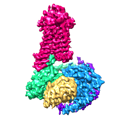 emd_20223.png emd_20223.png | 122.7 KB | ||
| Filedesc metadata | emd-20223.cif.gz | 6.8 KB | ||
| Archive directory |  http://ftp.pdbj.org/pub/emdb/structures/EMD-20223ftp://ftp.pdbj.org/pub/emdb/structures/EMD-20223 http://ftp.pdbj.org/pub/emdb/structures/EMD-20223ftp://ftp.pdbj.org/pub/emdb/structures/EMD-20223 | HTTPS FTP |
-Related structure data
| Related structure data |  6oyaMC  6oy9C C: citing same article ( M: atomic model generated by this map |
|---|---|
| Similar structure data |
-Links
| EMDB pages | EMDB (EBI/PDBe) / EMDataResource |
|---|---|
| Related items in Molecule of the Month |


-Map
| File | Download / File: emd_20223.map.gz / Format: CCP4 / Size: 52.7 MB / Type: IMAGE STORED AS FLOATING POINT NUMBER (4 BYTES) | ||||||||||||||||||||||||||||||||||||||||||||||||||||||||||||||||||||
|---|---|---|---|---|---|---|---|---|---|---|---|---|---|---|---|---|---|---|---|---|---|---|---|---|---|---|---|---|---|---|---|---|---|---|---|---|---|---|---|---|---|---|---|---|---|---|---|---|---|---|---|---|---|---|---|---|---|---|---|---|---|---|---|---|---|---|---|---|---|
| Annotation | Rhodopsin-Transducin-Nanobody Complex | ||||||||||||||||||||||||||||||||||||||||||||||||||||||||||||||||||||
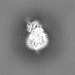
| Projections & slices | Image control
Images are generated by Spider. | ||||||||||||||||||||||||||||||||||||||||||||||||||||||||||||||||||||
| Voxel size | X=Y=Z: 1.06 Å | ||||||||||||||||||||||||||||||||||||||||||||||||||||||||||||||||||||
| Density |
| ||||||||||||||||||||||||||||||||||||||||||||||||||||||||||||||||||||
| Symmetry | Space group: 1 | ||||||||||||||||||||||||||||||||||||||||||||||||||||||||||||||||||||
| Details | EMDB XML:
CCP4 map header:
| ||||||||||||||||||||||||||||||||||||||||||||||||||||||||||||||||||||
 Z (Sec.)
 Y (Row.)
 X (Col.)


-Supplemental data
- Sample components
+Entire : Rhodopsin-Transducin-Nanobody Complex
+Supramolecule #1: Rhodopsin-Transducin-Nanobody Complex
+Supramolecule #2: Transducin
+Supramolecule #3: Rhodopsin
+Supramolecule #4: Nanobody
+Macromolecule #1: Gt-alpha/Gi1-alpha chimera

+Macromolecule #2: Guanine nucleotide-binding protein G(I)/G(S)/G(T) subunit beta-1
+Macromolecule #3: Guanine nucleotide-binding protein G(T) subunit gamma-T1
+Macromolecule #4: Camelid antibody VHH fragment
+Macromolecule #5: Rhodopsin
+Macromolecule #6: RETINAL

-Experimental details
-Structure determination
| Method | cryo EM |
|---|---|
 Processing Processing | single particle reconstruction |
| Aggregation state | particle |
-Sample preparation
| Buffer | pH: 7.5 |
|---|---|
| Grid | Model: Quantifoil R1.2/1.3 / Material: GOLD / Mesh: 200 / Pretreatment - Type: GLOW DISCHARGE / Pretreatment - Time: 80 sec. / Pretreatment - Atmosphere: AIR |
| Vitrification | Cryogen name: ETHANE / Chamber humidity: 100 % / Instrument: FEI VITROBOT MARK III Details: Blot for 1 second before plunging; avoid light as much as possible.. |
- Electron microscopy
| Microscope | FEI TITAN KRIOS |
|---|---|
| Specialist optics | Energy filter - Name: GIF Quantum LS / Energy filter - Slit width: 20 eV |
| Image recording | Film or detector model: GATAN K2 SUMMIT (4k x 4k) / Detector mode: COUNTING / Number real images: 3837 / Average electron dose: 40.0 e/Å2 |
| Electron beam | Acceleration voltage: 300 kV / Electron source:  FIELD EMISSION GUN FIELD EMISSION GUN |
| Electron optics | C2 aperture diameter: 50.0 µm / Illumination mode: FLOOD BEAM / Imaging mode: BRIGHT FIELD / Cs: 2.7 mm |
| Experimental equipment |  Model: Titan Krios / Image courtesy: FEI Company |
-Image processing
| Startup model | Type of model: EMDB MAP |
|---|---|
| Final reconstruction | Applied symmetry - Point group: C1 (asymmetric) / Resolution.type: BY AUTHOR / Resolution: 3.3 Å / Resolution method: FSC 0.143 CUT-OFF / Software - Name: cisTEM / Number images used: 309116 |
| Initial angle assignment | Type: PROJECTION MATCHING |
| Final angle assignment | Type: MAXIMUM LIKELIHOOD |
-Atomic model buiding 1
| Refinement | Protocol: BACKBONE TRACE |
|---|---|
| Output model | PDB-6oya: |
