 ムービー
ムービー コントローラー


+ データを開く

- 基本情報
基本情報
| 登録情報 | データベース: EMDB / ID: EMD-2020 | |||||||||
|---|---|---|---|---|---|---|---|---|---|---|
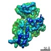
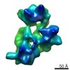
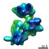
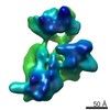
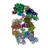
| タイトル | Structure of the E. coli 30S ribosomal subunit from a ksgA deletion strain in the conformation obtained at high magnesium concentrations | |||||||||
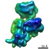
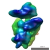
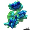
 マップデータ マップデータ | E. coli 30S ribosomal subunit from a ksgA deletion strain in the conformation obtained at high magnesium concentrations. | |||||||||
 試料 試料 |
| |||||||||
 キーワード キーワード | ribosome biogenesis / methyltransferase / KsgA / small ribosomal subunit | |||||||||
| 生物種 |  | |||||||||
| 手法 | 単粒子再構成法 / クライオ電子顕微鏡法 / 解像度: 17.7 Å | |||||||||
 データ登録者 データ登録者 | Boehringer D / O'Farrell HC / Rife JP / Ban N | |||||||||
 引用 引用 | ジャーナル: J Biol Chem / 年: 2012 タイトル: Structural insights into methyltransferase KsgA function in 30S ribosomal subunit biogenesis. 著者: Daniel Boehringer / Heather C O'Farrell / Jason P Rife / Nenad Ban /   要旨: The assembly of the ribosomal subunits is facilitated by ribosome biogenesis factors. The universally conserved methyltransferase KsgA modifies two adjacent adenosine residues in the 3'-terminal ...The assembly of the ribosomal subunits is facilitated by ribosome biogenesis factors. The universally conserved methyltransferase KsgA modifies two adjacent adenosine residues in the 3'-terminal helix 45 of the 16 S ribosomal RNA (rRNA). KsgA recognizes its substrate adenosine residues only in the context of a near mature 30S subunit and is required for the efficient processing of the rRNA termini during ribosome biogenesis. Here, we present the cryo-EM structure of KsgA bound to a nonmethylated 30S ribosomal subunit. The structure reveals that KsgA binds to the 30S platform with the catalytic N-terminal domain interacting with substrate adenosine residues in helix 45 and the C-terminal domain making extensive contacts to helix 27 and helix 24. KsgA excludes the penultimate rRNA helix 44 from adopting its position in the mature 30S subunit, blocking the formation of the decoding site and subunit joining. We suggest that the activation of methyltransferase activity and subsequent dissociation of KsgA control conformational changes in helix 44 required for final rRNA processing and translation initiation. | |||||||||
| 履歴 |
|
- 構造の表示
構造の表示
| ムービー |
 ムービービューア ムービービューア |
|---|---|
| 構造ビューア | EMマップ: SurfViewMolmilJmol/JSmol |
| 添付画像 |

- ダウンロードとリンク
-EMDBアーカイブ
| マップデータ | emd_2020.map.gz | 2.3 MB | EMDBマップデータ形式 | |
|---|---|---|---|---|
| ヘッダ (付随情報) | emd-2020-v30.xmlemd-2020.xml | 8.8 KB 8.8 KB | 表示 表示 | EMDBヘッダ |
| 画像 | 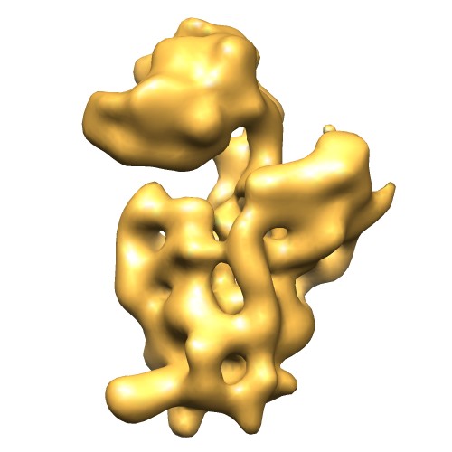 EMD-2020.jpg EMD-2020.jpg | 30.9 KB | ||
| アーカイブディレクトリ |  http://ftp.pdbj.org/pub/emdb/structures/EMD-2020ftp://ftp.pdbj.org/pub/emdb/structures/EMD-2020 http://ftp.pdbj.org/pub/emdb/structures/EMD-2020ftp://ftp.pdbj.org/pub/emdb/structures/EMD-2020 | HTTPS FTP |
-関連構造データ

-リンク
| EMDBのページ | EMDB (EBI/PDBe) / EMDataResource |
|---|---|
| 「今月の分子」の関連する項目 |

-マップ
| ファイル | ダウンロード / ファイル: emd_2020.map.gz / 形式: CCP4 / 大きさ: 2.5 MB / タイプ: IMAGE STORED AS FLOATING POINT NUMBER (4 BYTES) | ||||||||||||||||||||||||||||||||||||||||||||||||||||||||||||||||||||
|---|---|---|---|---|---|---|---|---|---|---|---|---|---|---|---|---|---|---|---|---|---|---|---|---|---|---|---|---|---|---|---|---|---|---|---|---|---|---|---|---|---|---|---|---|---|---|---|---|---|---|---|---|---|---|---|---|---|---|---|---|---|---|---|---|---|---|---|---|---|
| 注釈 | E. coli 30S ribosomal subunit from a ksgA deletion strain in the conformation obtained at high magnesium concentrations. | ||||||||||||||||||||||||||||||||||||||||||||||||||||||||||||||||||||
| 投影像・断面図 | 画像のコントロール
画像は Spider により作成 | ||||||||||||||||||||||||||||||||||||||||||||||||||||||||||||||||||||
| ボクセルのサイズ | X=Y=Z: 4.47 Å | ||||||||||||||||||||||||||||||||||||||||||||||||||||||||||||||||||||
| 密度 |
| ||||||||||||||||||||||||||||||||||||||||||||||||||||||||||||||||||||
| 対称性 | 空間群: 1 | ||||||||||||||||||||||||||||||||||||||||||||||||||||||||||||||||||||
| 詳細 | EMDB XML:
CCP4マップ ヘッダ情報:
| ||||||||||||||||||||||||||||||||||||||||||||||||||||||||||||||||||||
 Z (Sec.)
 Y (Row.)
 X (Col.)


-添付データ
- 試料の構成要素
-全体 : Structure of the E. coli 30S ribosomal subunit from a ksgA deleti...
| 全体 | 名称: Structure of the E. coli 30S ribosomal subunit from a ksgA deletion strain in the conformation obtained at high magnesium concentrations. |
|---|---|
| 要素 |
|
-超分子 #1000: Structure of the E. coli 30S ribosomal subunit from a ksgA deleti...
| 超分子 | 名称: Structure of the E. coli 30S ribosomal subunit from a ksgA deletion strain in the conformation obtained at high magnesium concentrations. タイプ: sample / ID: 1000 詳細: Translationally inactive 30S ribosomal subunits from a magnesium depleted sample were reactivated by incubation at high magnesium concentrations. 集合状態: Monomer / Number unique components: 1 |
|---|---|
| 分子量 | 実験値: 790 KDa / 理論値: 790 KDa |
-超分子 #1: E. coli 30S ribosomal subunit
| 超分子 | 名称: E. coli 30S ribosomal subunit / タイプ: complex / ID: 1 / Name.synonym: small ribosomal subunit 詳細: 30S ribosomal subunits lacking the methylation at A1518 and A1519 組換発現: Yes / Ribosome-details: ribosome-prokaryote: SSU 30S |
|---|---|
| 由来(天然) | 生物種: |
| 組換発現 | 生物種: |
| 分子量 | 実験値: 790 KDa / 理論値: 790 KDa |
-実験情報
-構造解析
| 手法 | クライオ電子顕微鏡法 |
|---|---|
 解析 解析 | 単粒子再構成法 |
| 試料の集合状態 | particle |
-試料調製
| 緩衝液 | pH: 7.6 詳細: 100 mM NH4Cl, 20 mM MgCl2, 20 mM HEPES/KOH pH 7.6, 3 mM DTT |
|---|---|
| グリッド | 詳細: Quantifoil 200 mesh copper |
| 凍結 | 凍結剤: ETHANE / 装置: OTHER |
- 電子顕微鏡法
| 顕微鏡 | FEI TECNAI 20 |
|---|---|
| 詳細 | Semi-automatic data acquisition with SerialEM script |
| 撮影 | カテゴリ: CCD フィルム・検出器のモデル: GATAN ULTRASCAN 4000 (4k x 4k) デジタル化 - サンプリング間隔: 15 µm / 平均電子線量: 20 e/Å2 / ビット/ピクセル: 16 |
| 電子線 | 加速電圧: 200 kV / 電子線源:  FIELD EMISSION GUN FIELD EMISSION GUN |
| 電子光学系 | 倍率(補正後): 82000 / 照射モード: SPOT SCAN / 撮影モード: BRIGHT FIELD / 最大 デフォーカス(公称値): 4.0 µm / 最小 デフォーカス(公称値): 2.0 µm / 倍率(公称値): 62000 |
| 試料ステージ | 試料ホルダー: Eucentric / 試料ホルダーモデル: GATAN LIQUID NITROGEN |
-画像解析
| CTF補正 | 詳細: Per image, ctffind3 |
|---|---|
| 最終 再構成 | 想定した対称性 - 点群: C1 (非対称) / アルゴリズム: OTHER / 解像度のタイプ: BY AUTHOR / 解像度: 17.7 Å / 解像度の算出法: FSC 0.5 CUT-OFF / ソフトウェア - 名称: Imagic-5,Spider / 使用した粒子像数: 15788 |
