 Movie
Movie Controller

[English] 日本語
 Yorodumi
- EMDB-5597: Structure of the immature 30S subunit from a Escherichia coli rim... -
+ Open data

- Basic information
| Entry | Database: EMDB / ID: EMD-5597 | |||||||||
|---|---|---|---|---|---|---|---|---|---|---|
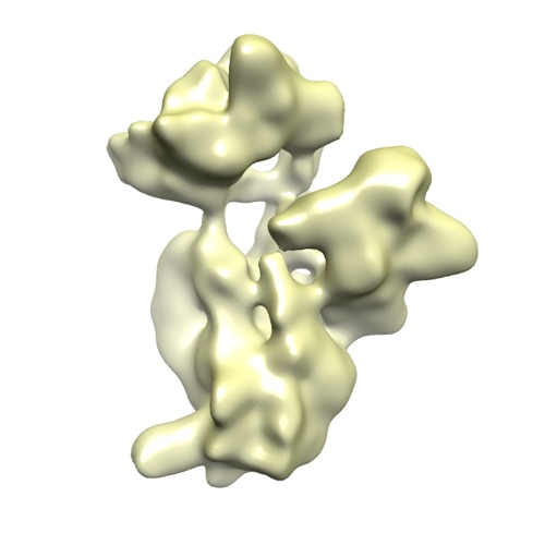
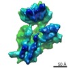
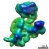
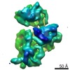
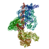
| Title | Structure of the immature 30S subunit from a Escherichia coli rimM null strain. Conformation 3. | |||||||||
 Map data Map data | Structure of the immature 30S subunit from a Escherichia coli rimM null strain. Conformation 3. | |||||||||
 Sample Sample |
| |||||||||
 Keywords Keywords | Ribosome assembly / 30S subunit / RimM protein | |||||||||
| Biological species |  | |||||||||
| Method | single particle reconstruction / cryo EM / Resolution: 17.0 Å | |||||||||
 Authors Authors | Leong V / Kent M / Jomaa A / Ortega J | |||||||||
 Citation Citation | Journal: RNA / Year: 2013 Title: Escherichia coli rimM and yjeQ null strains accumulate immature 30S subunits of similar structure and protein complement. Authors: Vivian Leong / Meredith Kent / Ahmad Jomaa / Joaquin Ortega /  Abstract: Assembly of the Escherichia coli 30S ribosomal subunits proceeds through multiple parallel pathways. The protein factors RimM, YjeQ, RbfA, and Era work in conjunction to assist at the late stages of ...Assembly of the Escherichia coli 30S ribosomal subunits proceeds through multiple parallel pathways. The protein factors RimM, YjeQ, RbfA, and Era work in conjunction to assist at the late stages of the maturation process of the small subunit. However, it is unclear how the functional interplay between these factors occurs in the context of multiple parallel pathways. To understand how these factors work together, we have characterized the immature 30S subunits that accumulate in ΔrimM cells and compared them with immature 30S subunits from a ΔyjeQ strain. The cryo-EM maps obtained from these particles showed that the densities representing helices 44 and 45 in the rRNA were partially missing, suggesting mobility of these motifs. These 30S subunits were also partially depleted in all tertiary ribosomal proteins, particularly those binding in the head domain. Using image classification, we identified four subpopulations of ΔrimM immature 30S subunits differing in the amount of missing density for helices 44 and 45, as well as the amount of density existing in these maps for the underrepresented proteins. The structural defects found in these immature subunits resembled those of the 30S subunits that accumulate in the ΔyjeQ strain. These findings are consistent with an "early convergency model" in which multiple parallel assembly pathways of the 30S subunit converge into a late assembly intermediate, as opposed to the mature state. Functionally related factors will bind to this intermediate to catalyze the last steps of maturation leading to the mature 30S subunit. | |||||||||
| History |
|
- Structure visualization
| Movie |
 Movie viewer Movie viewer |
|---|---|
| Structure viewer | EM map: SurfViewMolmilJmol/JSmol |
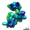
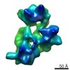
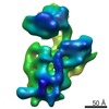
| Supplemental images |
- Downloads & links
-EMDB archive
| Map data | emd_5597.map.gz | 7.4 MB | EMDB map data format | |
|---|---|---|---|---|
| Header (meta data) | emd-5597-v30.xmlemd-5597.xml | 10.3 KB 10.3 KB | Display Display | EMDB header |
| Images | emd_5597.tif | 757.8 KB | ||
| Archive directory |  http://ftp.pdbj.org/pub/emdb/structures/EMD-5597ftp://ftp.pdbj.org/pub/emdb/structures/EMD-5597 http://ftp.pdbj.org/pub/emdb/structures/EMD-5597ftp://ftp.pdbj.org/pub/emdb/structures/EMD-5597 | HTTPS FTP |
-Related structure data

-Links
| EMDB pages | EMDB (EBI/PDBe) / EMDataResource |
|---|---|
| Related items in Molecule of the Month |

-Map
| File | Download / File: emd_5597.map.gz / Format: CCP4 / Size: 7.8 MB / Type: IMAGE STORED AS FLOATING POINT NUMBER (4 BYTES) | ||||||||||||||||||||||||||||||||||||||||||||||||||||||||||||||||||||
|---|---|---|---|---|---|---|---|---|---|---|---|---|---|---|---|---|---|---|---|---|---|---|---|---|---|---|---|---|---|---|---|---|---|---|---|---|---|---|---|---|---|---|---|---|---|---|---|---|---|---|---|---|---|---|---|---|---|---|---|---|---|---|---|---|---|---|---|---|---|
| Annotation | Structure of the immature 30S subunit from a Escherichia coli rimM null strain. Conformation 3. | ||||||||||||||||||||||||||||||||||||||||||||||||||||||||||||||||||||
| Projections & slices | Image control
Images are generated by Spider. | ||||||||||||||||||||||||||||||||||||||||||||||||||||||||||||||||||||
| Voxel size | X=Y=Z: 2.54 Å | ||||||||||||||||||||||||||||||||||||||||||||||||||||||||||||||||||||
| Density |
| ||||||||||||||||||||||||||||||||||||||||||||||||||||||||||||||||||||
| Symmetry | Space group: 1 | ||||||||||||||||||||||||||||||||||||||||||||||||||||||||||||||||||||
| Details | EMDB XML:
CCP4 map header:
| ||||||||||||||||||||||||||||||||||||||||||||||||||||||||||||||||||||
 Z (Sec.)
 Y (Row.)
 X (Col.)


-Supplemental data
- Sample components
-Entire : Structure of an immature 30S subunit from an E. coli rimM null st...
| Entire | Name: Structure of an immature 30S subunit from an E. coli rimM null strain. Conformation 3 |
|---|---|
| Components |
|
-Supramolecule #1000: Structure of an immature 30S subunit from an E. coli rimM null st...
| Supramolecule | Name: Structure of an immature 30S subunit from an E. coli rimM null strain. Conformation 3 type: sample / ID: 1000 / Oligomeric state: monomer / Number unique components: 1 |
|---|---|
| Molecular weight | Experimental: 930 KDa / Theoretical: 930 KDa |
-Supramolecule #1: ribosome 30S subunit
| Supramolecule | Name: ribosome 30S subunit / type: complex / ID: 1 / Recombinant expression: No / Database: NCBI / Ribosome-details: ribosome-prokaryote: SSU 30S, PSR16s |
|---|---|
| Source (natural) | Organism: |
| Molecular weight | Experimental: 930 KDa / Theoretical: 930 KDa |
-Experimental details
-Structure determination
| Method | cryo EM |
|---|---|
 Processing Processing | single particle reconstruction |
| Aggregation state | particle |
-Sample preparation
| Buffer | pH: 7.5 Details: 10 mM Tris-HCl, pH 7.5, 10 mM Mg acetate, 60 mM NH4Cl and 3 mM 2-mercaptoethanol |
|---|---|
| Grid | Details: 400 mesh cupped grid with thin carbon support, glow discharged in air |
| Vitrification | Cryogen name: ETHANE / Chamber humidity: 100 % / Chamber temperature: 77 K / Instrument: FEI VITROBOT MARK III / Method: Blot twice for 7 seconds each. |
- Electron microscopy
| Microscope | JEOL 2010F |
|---|---|
| Temperature | Average: 97 K |
| Date | Jul 10, 2011 |
| Image recording | Category: FILM / Film or detector model: KODAK SO-163 FILM / Digitization - Scanner: NIKON SUPER COOLSCAN 9000 / Digitization - Sampling interval: 6.35 µm / Number real images: 250 / Average electron dose: 15 e/Å2 / Bits/pixel: 8 |
| Tilt angle min | 0 |
| Tilt angle max | 0 |
| Electron beam | Acceleration voltage: 200 kV / Electron source:  FIELD EMISSION GUN FIELD EMISSION GUN |
| Electron optics | Illumination mode: FLOOD BEAM / Imaging mode: BRIGHT FIELD / Cs: 1 mm / Nominal defocus max: 4.0 µm / Nominal defocus min: 1.5 µm / Nominal magnification: 50000 |
| Sample stage | Specimen holder: Liquid nitrogen cooled / Specimen holder model: GATAN LIQUID NITROGEN |
-Image processing
| Details | Reconstruction obtained with Xmipp |
|---|---|
| CTF correction | Details: CTFFIND |
| Final reconstruction | Algorithm: OTHER / Resolution.type: BY AUTHOR / Resolution: 17.0 Å / Resolution method: FSC 0.5 CUT-OFF / Software - Name: Xmipp / Number images used: 23275 |
