Movie
Movie Controller


+ Open data

- Basic information
| Entry |  | |||||||||
|---|---|---|---|---|---|---|---|---|---|---|
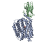
| Title | SPNS2:sfGFP hetero dimer assembled by Di-Gluebody | |||||||||
 Map data Map data | SPNS2:sfGFP hetero dimer assembled by Di-Gluebody | |||||||||
 Sample Sample |
| |||||||||
 Keywords Keywords | SPNS2 / Di-Gluebody / sfGFP / MEMBRANE PROTEIN | |||||||||
| Function / homology |  Function and homology information Function and homology informationregulation of eye pigmentation / regulation of humoral immune response / regulation of T cell migration / sphingolipid intramembrane carrier activity / lymphocyte migration / Sphingolipid de novo biosynthesis / sphingolipid biosynthetic process / sphingosine-1-phosphate receptor signaling pathway / lipid transport / B cell homeostasis ...regulation of eye pigmentation / regulation of humoral immune response / regulation of T cell migration / sphingolipid intramembrane carrier activity / lymphocyte migration / Sphingolipid de novo biosynthesis / sphingolipid biosynthetic process / sphingosine-1-phosphate receptor signaling pathway / lipid transport / B cell homeostasis / T cell homeostasis / lymph node development / transmembrane transporter activity / bioluminescence / generation of precursor metabolites and energy / sensory perception of sound / bone development / endosome membrane / membrane / plasma membrane Similarity search - Function | |||||||||
| Biological species |   Aequorea victoria (jellyfish) / Aequorea victoria (jellyfish) /   Homo sapiens (human) Homo sapiens (human) | |||||||||
| Method | single particle reconstruction / cryo EM / Resolution: 3.9 Å | |||||||||
 Authors Authors | Yi G / Ye M / Mamalis D / Li H / Sauer DB / von Delft F / Davis BG / Gilbert RJC | |||||||||
| Funding support |  United Kingdom, 1 items United Kingdom, 1 items
| |||||||||
 Citation Citation | Journal: Nat Chem Biol / Year: 2026 Title: Covalently constrained 'Di-Gembodies' enable parallel structure solutions by cryo-EM. Authors: Gangshun Yi / Dimitrios Mamalis / Mingda Ye / Loic Carrique / Michael Fairhead / Huanyu Li / Katharina L Duerr / Peijun Zhang / David B Sauer / Frank von Delft / Benjamin G Davis / Robert J C Gilbert /  Abstract: Whilst cryo-electron microscopy(cryo-EM) has become a routine methodology in structural biology, obtaining high-resolution cryo-EM structures of small proteins (<100 kDa) and increasing overall throughput remain challenging. One approach to augment protein size and improve particle alignment involves the use of binding proteins or protein-based scaffolds. However, a given imaging scaffold or linking module may prove inadequate for structure solution and availability of such scaffolds remains limited. Here, we describe a strategy that exploits covalent dimerization of nanobodies to trap an engineered, predisposed nanobody-to-nanobody interface, giving Di-Gembodies as modular constructs created in homomeric and heteromeric forms. By exploiting side-chain-to-side-chain assembly, they can simultaneously display two copies of the same or two distinct proteins through a subunit interface that provides sufficient constraint required for cryo-EM structure determination. We validate this method with multiple soluble and membrane structural targets, down to 14 kDa, demonstrating a flexible and scalable platform for expanded protein structure determination. | |||||||||
| History |
|
- Structure visualization
| Supplemental images |
|---|

- Downloads & links
-EMDB archive
| Map data | emd_19338.map.gz | 154.3 MB | EMDB map data format | |
|---|---|---|---|---|
| Header (meta data) | emd-19338-v30.xmlemd-19338.xml | 29.5 KB 29.5 KB | Display Display | EMDB header |
| FSC (resolution estimation) | emd_19338_fsc.xml | 11.6 KB | Display | FSC data file |
| Images |  emd_19338.png emd_19338.png | 67.1 KB | ||
| Filedesc metadata | emd-19338.cif.gz | 7.6 KB | ||
| Others | emd_19338_half_map_1.map.gzemd_19338_half_map_2.map.gz | 151.6 MB 151.6 MB | ||
| Archive directory |  http://ftp.pdbj.org/pub/emdb/structures/EMD-19338ftp://ftp.pdbj.org/pub/emdb/structures/EMD-19338 http://ftp.pdbj.org/pub/emdb/structures/EMD-19338ftp://ftp.pdbj.org/pub/emdb/structures/EMD-19338 | HTTPS FTP |
-Related structure data
| Related structure data |  8rlcMC  8rl5C  8rl6C  8rl7C  8rl8C  8rl9C  8rlaC  8rlbC  8rldC  8rleC 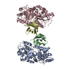 9fgvC  9fgxC  9fgyC  9fkqC M: atomic model generated by this map C: citing same article ( |
|---|---|
| Similar structure data |


-Links
| EMDB pages | EMDB (EBI/PDBe) / EMDataResource |
|---|---|
| Related items in Molecule of the Month |


-Map
| File | Download / File: emd_19338.map.gz / Format: CCP4 / Size: 163.6 MB / Type: IMAGE STORED AS FLOATING POINT NUMBER (4 BYTES) | ||||||||||||||||||||||||||||||||||||
|---|---|---|---|---|---|---|---|---|---|---|---|---|---|---|---|---|---|---|---|---|---|---|---|---|---|---|---|---|---|---|---|---|---|---|---|---|---|
| Annotation | SPNS2:sfGFP hetero dimer assembled by Di-Gluebody | ||||||||||||||||||||||||||||||||||||
| Projections & slices | Image control
Images are generated by Spider. | ||||||||||||||||||||||||||||||||||||
| Voxel size | X=Y=Z: 0.932 Å | ||||||||||||||||||||||||||||||||||||
| Density |
| ||||||||||||||||||||||||||||||||||||
| Symmetry | Space group: 1 | ||||||||||||||||||||||||||||||||||||
| Details | EMDB XML:
|
 Z (Sec.)
 Y (Row.)
 X (Col.)


-Supplemental data
-Half map: half map B
| File | emd_19338_half_map_1.map | ||||||||||||
|---|---|---|---|---|---|---|---|---|---|---|---|---|---|
| Annotation | half map B | ||||||||||||
| Projections & Slices |
| ||||||||||||
| Density Histograms |


-Half map: half map A
| File | emd_19338_half_map_2.map | ||||||||||||
|---|---|---|---|---|---|---|---|---|---|---|---|---|---|
| Annotation | half map A | ||||||||||||
| Projections & Slices |
| ||||||||||||
| Density Histograms |


- Sample components
-Entire : SPNS2:sfGFP heterodimer assembled by Di-Gluebody
| Entire | Name: SPNS2:sfGFP heterodimer assembled by Di-Gluebody |
|---|---|
| Components |
|
-Supramolecule #1: SPNS2:sfGFP heterodimer assembled by Di-Gluebody
| Supramolecule | Name: SPNS2:sfGFP heterodimer assembled by Di-Gluebody / type: complex / ID: 1 / Parent: 0 / Macromolecule list: #1-#4 Details: GbEnhancer and GbC4 were assembled via a disulfide to form a hetero Di-Gluebody |
|---|---|
| Molecular weight | Theoretical: 112 KDa |
-Supramolecule #2: Green fluorescent protein
| Supramolecule | Name: Green fluorescent protein / type: complex / ID: 2 / Parent: 1 / Macromolecule list: #1 / Details: Green fluorescent protein |
|---|---|
| Source (natural) | Organism: Aequorea victoria (jellyfish) |
-Supramolecule #3: Di-Gluebody
| Supramolecule | Name: Di-Gluebody / type: complex / ID: 3 / Parent: 1 / Macromolecule list: #2-#3 / Details: Gluebody GbEnhancer and Gluebody GbC4 |
|---|---|
| Source (natural) | Organism: |
-Supramolecule #4: Sphingosine-1-phosphate transporter SPNS2
| Supramolecule | Name: Sphingosine-1-phosphate transporter SPNS2 / type: complex / ID: 4 / Parent: 1 / Macromolecule list: #4 / Details: Sphingosine-1-phosphate transporter SPNS2 |
|---|---|
| Source (natural) | Organism: Homo sapiens (human) |
-Macromolecule #1: Green fluorescent protein
| Macromolecule | Name: Green fluorescent protein / type: protein_or_peptide / ID: 1 / Number of copies: 1 / Enantiomer: LEVO |
|---|---|
| Source (natural) | Organism: Aequorea victoria (jellyfish) |
| Molecular weight | Theoretical: 26.81923 KDa |
| Recombinant expression | Organism:  |
| Sequence | String: MSKGEELFTG VVPILVELDG DVNGHKFSVR GEGEGDATNG KLTLKFICTT GKLPVPWPTL VTTLTYGVQC FSRYPDHMKR HDFFKSAMP EGYVQERTIS FKDDGTYKTR AEVKFEGDTL VNRIELKGID FKEDGNILGH KLEYNFNSHN VYITADKQKN G IKANFKIR ...String: MSKGEELFTG VVPILVELDG DVNGHKFSVR GEGEGDATNG KLTLKFICTT GKLPVPWPTL VTTLTYGVQC FSRYPDHMKR HDFFKSAMP EGYVQERTIS FKDDGTYKTR AEVKFEGDTL VNRIELKGID FKEDGNILGH KLEYNFNSHN VYITADKQKN G IKANFKIR HNVEDGSVQL ADHYQQNTPI GDGPVLLPDN HYLSTQSVLS KDPNEKRDHM VLLEFVTAAG ITHGMDELYK UniProtKB: Green fluorescent protein |
-Macromolecule #2: Gluebody GbEnhancer
| Macromolecule | Name: Gluebody GbEnhancer / type: protein_or_peptide / ID: 2 / Number of copies: 1 / Enantiomer: LEVO |
|---|---|
| Source (natural) | Organism: |
| Molecular weight | Theoretical: 12.602062 KDa |
| Recombinant expression | Organism: |
| Sequence | String: QVQLVENGGA CVKPGGSLRL SCAASGFPVN RYSMRWYRQA PGKEREWVAG MSSAGDRSSY EDSVKGRFTI SRDDARNTVY LQMNSLKPE DTAVYYCNVN VGFEYWGQGT QVMV |
-Macromolecule #3: Gluebody GbC4
| Macromolecule | Name: Gluebody GbC4 / type: protein_or_peptide / ID: 3 / Number of copies: 1 / Enantiomer: LEVO |
|---|---|
| Source (natural) | Organism: |
| Molecular weight | Theoretical: 12.982609 KDa |
| Recombinant expression | Organism: |
| Sequence | String: SQGQLVENGG GCVKAGGSLR LSCAASQGTL SNLVTGWFRR APGKEREFVA NIGRDGLTVY SNSVKGRFTI SRDRAKNTVY LQMDSLKPE DTAVYYCAGR LSRFPGEYDY WSKGTPVMVS |
-Macromolecule #4: Sphingosine-1-phosphate transporter SPNS2
| Macromolecule | Name: Sphingosine-1-phosphate transporter SPNS2 / type: protein_or_peptide / ID: 4 / Number of copies: 1 / Enantiomer: LEVO |
|---|---|
| Source (natural) | Organism: Homo sapiens (human) |
| Molecular weight | Theoretical: 58.962211 KDa |
| Recombinant expression | Organism: Homo sapiens (human) |
| Sequence | String: MMCLECASAA AGGAEEEEAD AERRRRRRGA QRGAGGSGCC GARGAGGAGV SAAGDEVQTL SGSVRRAPTG PPGTPGTPGC AATAKGPGA QQPKPASLGR GRGAAAAILS LGNVLNYLDR YTVAGVLLDI QQHFGVKDRG AGLLQSVFIC SFMVAAPIFG Y LGDRFNRK ...String: MMCLECASAA AGGAEEEEAD AERRRRRRGA QRGAGGSGCC GARGAGGAGV SAAGDEVQTL SGSVRRAPTG PPGTPGTPGC AATAKGPGA QQPKPASLGR GRGAAAAILS LGNVLNYLDR YTVAGVLLDI QQHFGVKDRG AGLLQSVFIC SFMVAAPIFG Y LGDRFNRK VILSCGIFFW SAVTFSSSFI PQQYFWLLVL SRGLVGIGEA SYSTIAPTII GDLFTKNTRT LMLSVFYFAI PL GSGLGYI TGSSVKQAAG DWHWALRVSP VLGMITGTLI LILVPATKRG HADQLGDQLK ARTSWLRDMK ALIRNRSYVF SSL ATSAVS FATGALGMWI PLYLHRAQVV QKTAETCNSP PCGAKDSLIF GAITCFTGFL GVVTGAGATR WCRLKTQRAD PLVC AVGML GSAIFICLIF VAAKSSIVGA YICIFVGETL LFSNWAITAD ILMYVVIPTR RATAVALQSF TSHLLGDAGS PYLIG FISD LIRQSTKDSP LWEFLSLGYA LMLCPFVVVL GGMFFLATAL FFVSDRARAE QQVNQLAMPP ASVKVAENLY FQ UniProtKB: Sphingosine-1-phosphate transporter SPNS2 |
-Macromolecule #5: DODECYL-BETA-D-MALTOSIDE
| Macromolecule | Name: DODECYL-BETA-D-MALTOSIDE / type: ligand / ID: 5 / Number of copies: 1 / Formula: LMT |
|---|---|
| Molecular weight | Theoretical: 510.615 Da |
| Chemical component information |  ChemComp-LMT: |
-Experimental details
-Structure determination
| Method | cryo EM |
|---|---|
 Processing Processing | single particle reconstruction |
| Aggregation state | particle |
-Sample preparation
| Concentration | 4.2 mg/mL | ||||||||||||
|---|---|---|---|---|---|---|---|---|---|---|---|---|---|
| Buffer | pH: 7.5 Component:
| ||||||||||||
| Grid | Model: Quantifoil R1.2/1.3 / Material: COPPER / Mesh: 300 / Pretreatment - Type: GLOW DISCHARGE | ||||||||||||
| Vitrification | Cryogen name: ETHANE |
- Electron microscopy
| Microscope | FEI TITAN KRIOS |
|---|---|
| Image recording | Film or detector model: GATAN K3 (6k x 4k) / Number real images: 8674 / Average electron dose: 50.0 e/Å2 |
| Electron beam | Acceleration voltage: 300 kV / Electron source:  FIELD EMISSION GUN FIELD EMISSION GUN |
| Electron optics | Illumination mode: FLOOD BEAM / Imaging mode: BRIGHT FIELD / Nominal defocus max: 2.5 µm / Nominal defocus min: 1.5 µm |
| Experimental equipment |  Model: Titan Krios / Image courtesy: FEI Company |