 Movie
Movie Controller


+ Open data

- Basic information
| Entry |  | |||||||||
|---|---|---|---|---|---|---|---|---|---|---|
| Title | RECQL5:sfGFP hetero dimer assembled by Di-Gluebody | |||||||||
 Map data Map data | EMhancer: RECQL5:sfGFP hetero dimer complex assembled by Di-Gluebody | |||||||||
 Sample Sample |
| |||||||||
 Keywords Keywords | DNA helicase / Di-Gluebody / GFP / HYDROLASE | |||||||||
| Function / homology |  Function and homology information Function and homology informationmitotic DNA-templated DNA replication / chromosome separation / four-way junction helicase activity / cellular response to camptothecin / replication-born double-strand break repair via sister chromatid exchange / transcription preinitiation complex / DNA metabolic process / 3'-5' DNA helicase activity / RNA polymerase II complex binding / DNA 3'-5' helicase ...mitotic DNA-templated DNA replication / chromosome separation / four-way junction helicase activity / cellular response to camptothecin / replication-born double-strand break repair via sister chromatid exchange / transcription preinitiation complex / DNA metabolic process / 3'-5' DNA helicase activity / RNA polymerase II complex binding / DNA 3'-5' helicase / negative regulation of transcription elongation by RNA polymerase II / negative regulation of double-strand break repair via homologous recombination / bioluminescence / replication fork / DNA helicase activity / helicase activity / generation of precursor metabolites and energy / double-strand break repair via homologous recombination / cellular response to xenobiotic stimulus / mitotic cell cycle / chromosome / DNA replication / cell division / DNA repair / ATP hydrolysis activity / DNA binding / nucleoplasm / ATP binding / metal ion binding / identical protein binding / nucleus / cytoplasm / cytosol Similarity search - Function | |||||||||
| Biological species |   Aequorea victoria (jellyfish) / Aequorea victoria (jellyfish) /   Homo sapiens (human) Homo sapiens (human) | |||||||||
| Method | single particle reconstruction / cryo EM / Resolution: 3.22 Å | |||||||||
 Authors Authors | Yi G / Ye M / Mamalis D / Fairhead M / Sauer DB / von Delft F / Davis BG / Gilbert RJC | |||||||||
| Funding support |  United Kingdom, 1 items United Kingdom, 1 items
| |||||||||
 Citation Citation | Journal: Nat Chem Biol / Year: 2026 Title: Covalently constrained 'Di-Gembodies' enable parallel structure solutions by cryo-EM. Authors: Gangshun Yi / Dimitrios Mamalis / Mingda Ye / Loic Carrique / Michael Fairhead / Huanyu Li / Katharina L Duerr / Peijun Zhang / David B Sauer / Frank von Delft / Benjamin G Davis / Robert J C Gilbert /  Abstract: Whilst cryo-electron microscopy(cryo-EM) has become a routine methodology in structural biology, obtaining high-resolution cryo-EM structures of small proteins (<100 kDa) and increasing overall throughput remain challenging. One approach to augment protein size and improve particle alignment involves the use of binding proteins or protein-based scaffolds. However, a given imaging scaffold or linking module may prove inadequate for structure solution and availability of such scaffolds remains limited. Here, we describe a strategy that exploits covalent dimerization of nanobodies to trap an engineered, predisposed nanobody-to-nanobody interface, giving Di-Gembodies as modular constructs created in homomeric and heteromeric forms. By exploiting side-chain-to-side-chain assembly, they can simultaneously display two copies of the same or two distinct proteins through a subunit interface that provides sufficient constraint required for cryo-EM structure determination. We validate this method with multiple soluble and membrane structural targets, down to 14 kDa, demonstrating a flexible and scalable platform for expanded protein structure determination. | |||||||||
| History |
|
- Structure visualization
| Supplemental images |
|---|

- Downloads & links
-EMDB archive
| Map data | emd_19335.map.gz | 165.1 MB | EMDB map data format | |
|---|---|---|---|---|
| Header (meta data) | emd-19335-v30.xmlemd-19335.xml | 31.4 KB 31.4 KB | Display Display | EMDB header |
| FSC (resolution estimation) | emd_19335_fsc.xml | 11.9 KB | Display | FSC data file |
| Images |  emd_19335.png emd_19335.png | 68.7 KB | ||
| Filedesc metadata | emd-19335.cif.gz | 7.5 KB | ||
| Others | emd_19335_additional_1.map.gzemd_19335_half_map_1.map.gzemd_19335_half_map_2.map.gz | 167.8 MB 165 MB 165 MB | ||
| Archive directory |  http://ftp.pdbj.org/pub/emdb/structures/EMD-19335ftp://ftp.pdbj.org/pub/emdb/structures/EMD-19335 http://ftp.pdbj.org/pub/emdb/structures/EMD-19335ftp://ftp.pdbj.org/pub/emdb/structures/EMD-19335 | HTTPS FTP |
-Related structure data
| Related structure data |  8rl9MC  8rl5C  8rl6C  8rl7C  8rl8C  8rlaC  8rlbC  8rlcC  8rldC  8rleC 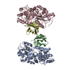 9fgvC  9fgxC  9fgyC  9fkqC M: atomic model generated by this map C: citing same article ( |
|---|---|
| Similar structure data |


-Links
| EMDB pages | EMDB (EBI/PDBe) / EMDataResource |
|---|---|
| Related items in Molecule of the Month |


-Map
| File | Download / File: emd_19335.map.gz / Format: CCP4 / Size: 178 MB / Type: IMAGE STORED AS FLOATING POINT NUMBER (4 BYTES) | ||||||||||||||||||||||||||||||||||||
|---|---|---|---|---|---|---|---|---|---|---|---|---|---|---|---|---|---|---|---|---|---|---|---|---|---|---|---|---|---|---|---|---|---|---|---|---|---|
| Annotation | EMhancer: RECQL5:sfGFP hetero dimer complex assembled by Di-Gluebody | ||||||||||||||||||||||||||||||||||||
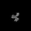
| Projections & slices | Image control
Images are generated by Spider. | ||||||||||||||||||||||||||||||||||||
| Voxel size | X=Y=Z: 0.83 Å | ||||||||||||||||||||||||||||||||||||
| Density |
| ||||||||||||||||||||||||||||||||||||
| Symmetry | Space group: 1 | ||||||||||||||||||||||||||||||||||||
| Details | EMDB XML:
|
 Z (Sec.)
 Y (Row.)
 X (Col.)


-Supplemental data
-Additional map: RECQL5:sfGFP hetero dimer complex assembled by Di-Gluebody
| File | emd_19335_additional_1.map | ||||||||||||
|---|---|---|---|---|---|---|---|---|---|---|---|---|---|
| Annotation | RECQL5:sfGFP hetero dimer complex assembled by Di-Gluebody | ||||||||||||
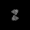
| Projections & Slices |
| ||||||||||||
| Density Histograms |


-Half map: half map B
| File | emd_19335_half_map_1.map | ||||||||||||
|---|---|---|---|---|---|---|---|---|---|---|---|---|---|
| Annotation | half map B | ||||||||||||
| Projections & Slices |
| ||||||||||||
| Density Histograms |


-Half map: half map A
| File | emd_19335_half_map_2.map | ||||||||||||
|---|---|---|---|---|---|---|---|---|---|---|---|---|---|
| Annotation | half map A | ||||||||||||
| Projections & Slices |
| ||||||||||||
| Density Histograms |


- Sample components
-Entire : RECQL5:sfGFP heterodimer assembled by Di-Gluebody
| Entire | Name: RECQL5:sfGFP heterodimer assembled by Di-Gluebody |
|---|---|
| Components |
|
-Supramolecule #1: RECQL5:sfGFP heterodimer assembled by Di-Gluebody
| Supramolecule | Name: RECQL5:sfGFP heterodimer assembled by Di-Gluebody / type: complex / ID: 1 / Parent: 0 / Macromolecule list: #1-#4 Details: GbEnhancer and G5-006 were assembled via a disulfide to form a hetero Di-Gluebody |
|---|---|
| Molecular weight | Theoretical: 103 KDa |
-Supramolecule #2: Green fluorescent protein
| Supramolecule | Name: Green fluorescent protein / type: complex / ID: 2 / Parent: 1 / Macromolecule list: #1 / Details: Green fluorescent protein |
|---|---|
| Source (natural) | Organism: Aequorea victoria (jellyfish) |
-Supramolecule #3: Di-Gluebody
| Supramolecule | Name: Di-Gluebody / type: complex / ID: 3 / Parent: 1 / Macromolecule list: #2-#3 / Details: Gluebody GbEnhancer and Gluebody G5-006 |
|---|---|
| Source (natural) | Organism: |
-Supramolecule #4: ATP-dependent DNA helicase Q5
| Supramolecule | Name: ATP-dependent DNA helicase Q5 / type: complex / ID: 4 / Parent: 1 / Macromolecule list: #4 / Details: ATP-dependent DNA helicase Q5 |
|---|---|
| Source (natural) | Organism: Homo sapiens (human) |
-Macromolecule #1: Green fluorescent protein
| Macromolecule | Name: Green fluorescent protein / type: protein_or_peptide / ID: 1 / Number of copies: 1 / Enantiomer: LEVO |
|---|---|
| Source (natural) | Organism: Aequorea victoria (jellyfish) |
| Molecular weight | Theoretical: 26.81923 KDa |
| Recombinant expression | Organism:  |
| Sequence | String: MSKGEELFTG VVPILVELDG DVNGHKFSVR GEGEGDATNG KLTLKFICTT GKLPVPWPTL VTTLTYGVQC FSRYPDHMKR HDFFKSAMP EGYVQERTIS FKDDGTYKTR AEVKFEGDTL VNRIELKGID FKEDGNILGH KLEYNFNSHN VYITADKQKN G IKANFKIR ...String: MSKGEELFTG VVPILVELDG DVNGHKFSVR GEGEGDATNG KLTLKFICTT GKLPVPWPTL VTTLTYGVQC FSRYPDHMKR HDFFKSAMP EGYVQERTIS FKDDGTYKTR AEVKFEGDTL VNRIELKGID FKEDGNILGH KLEYNFNSHN VYITADKQKN G IKANFKIR HNVEDGSVQL ADHYQQNTPI GDGPVLLPDN HYLSTQSVLS KDPNEKRDHM VLLEFVTAAG ITHGMDELYK UniProtKB: Green fluorescent protein |
-Macromolecule #2: Gluebody GbEnhancer
| Macromolecule | Name: Gluebody GbEnhancer / type: protein_or_peptide / ID: 2 / Number of copies: 1 / Enantiomer: LEVO |
|---|---|
| Source (natural) | Organism: |
| Molecular weight | Theoretical: 12.689139 KDa |
| Recombinant expression | Organism: |
| Sequence | String: QVQLVENGGA CVKPGGSLRL SCAASGFPVN RYSMRWYRQA PGKEREWVAG MSSAGDRSSY EDSVKGRFTI SRDDARNTVY LQMNSLKPE DTAVYYCNVN VGFEYWGQGT QVMVS |
-Macromolecule #3: Gluebody G5-006
| Macromolecule | Name: Gluebody G5-006 / type: protein_or_peptide / ID: 3 / Number of copies: 1 / Enantiomer: LEVO |
|---|---|
| Source (natural) | Organism: |
| Molecular weight | Theoretical: 13.775173 KDa |
| Recombinant expression | Organism: |
| Sequence | String: SMAQVQLVEN GGGCVKAGGS LRLSCAASGS IFSINRMTWY RQAPGKEREW VAAITSGGST NYADSVKGRF TISRDNAENT VYLQMNSLK PEDTAVYYCE AYGTYTLAPT GEGEYDDYWG QGTQVMVS |
-Macromolecule #4: ATP-dependent DNA helicase Q5
| Macromolecule | Name: ATP-dependent DNA helicase Q5 / type: protein_or_peptide / ID: 4 / Number of copies: 1 / Enantiomer: LEVO / EC number: DNA helicase |
|---|---|
| Source (natural) | Organism: Homo sapiens (human) |
| Molecular weight | Theoretical: 49.20382 KDa |
| Recombinant expression | Organism: |
| Sequence | String: PERRVRSTLK KVFGFDSFKT PLQESATMAV VKGNKDVFVC MPTGAGKSLC YQLPALLAKG ITIVVSPLIA LIQDQVDHLL TLKVRVSSL NSKLSAQERK ELLADLEREK PQTKILYITP EMAASSSFQP TLNSLVSRHL LSYLVVDEAH CVSQWGHDFR P DYLRLGAL ...String: PERRVRSTLK KVFGFDSFKT PLQESATMAV VKGNKDVFVC MPTGAGKSLC YQLPALLAKG ITIVVSPLIA LIQDQVDHLL TLKVRVSSL NSKLSAQERK ELLADLEREK PQTKILYITP EMAASSSFQP TLNSLVSRHL LSYLVVDEAH CVSQWGHDFR P DYLRLGAL RSRLGHAPCV ALTATATPQV QEDVFAALHL KKPVAIFKTP CFRANLFYDV QFKELISDPY GNLKDFCLKA LG QEADKGL SGCGIVYCRT REACEQLAIE LSCRGVNAKA YHAGLKASER TLVQNDWMEE KVPVIVATIS FGMGVDKANV RFV AHWNIA KSMAGYYQES GRAGRDGKPS WCRLYYSRND RDQVSFLIRK EVAKLQEKRG NKASDKATIM AFDALVTFCE ELGC RHAAI AKYFGDALPA CAKGCDHCQN PTAVRRRLEA LERSSSW UniProtKB: ATP-dependent DNA helicase Q5 |
-Macromolecule #5: ZINC ION
| Macromolecule | Name: ZINC ION / type: ligand / ID: 5 / Number of copies: 1 / Formula: ZN |
|---|---|
| Molecular weight | Theoretical: 65.409 Da |
-Experimental details
-Structure determination
| Method | cryo EM |
|---|---|
 Processing Processing | single particle reconstruction |
| Aggregation state | particle |
-Sample preparation
| Concentration | 1.1 mg/mL | |||||||||
|---|---|---|---|---|---|---|---|---|---|---|
| Buffer | pH: 7.5 Component:
| |||||||||
| Grid | Model: C-flat-2/1 / Material: GOLD / Mesh: 200 / Pretreatment - Type: GLOW DISCHARGE | |||||||||
| Vitrification | Cryogen name: ETHANE |
- Electron microscopy
| Microscope | FEI TITAN KRIOS |
|---|---|
| Image recording | Film or detector model: GATAN K3 (6k x 4k) / Number real images: 10102 / Average electron dose: 42.0 e/Å2 |
| Electron beam | Acceleration voltage: 300 kV / Electron source:  FIELD EMISSION GUN FIELD EMISSION GUN |
| Electron optics | Illumination mode: FLOOD BEAM / Imaging mode: BRIGHT FIELD / Nominal defocus max: 2.5 µm / Nominal defocus min: 1.5 µm |
| Experimental equipment |  Model: Titan Krios / Image courtesy: FEI Company |


