 Movie
Movie Controller

[English] 日本語
 Yorodumi
- PDB-9fgx: Cryo-EM structure of Lysozyme homo-dimer assembled by homo Di-Gluebody -
+ Open data

- Basic information
| Entry | Database: PDB / ID: 9fgx | ||||||||||||||||||||||||
|---|---|---|---|---|---|---|---|---|---|---|---|---|---|---|---|---|---|---|---|---|---|---|---|---|---|
| Title | Cryo-EM structure of Lysozyme homo-dimer assembled by homo Di-Gluebody | ||||||||||||||||||||||||
 Components Components |
| ||||||||||||||||||||||||
 Keywords Keywords | PROTEIN BINDING / Gluebody / Nanobody / cryo-EM SPA / small protein | ||||||||||||||||||||||||
| Function / homology |  Function and homology information Function and homology informationLactose synthesis / Antimicrobial peptides / Neutrophil degranulation / beta-N-acetylglucosaminidase activity / cell wall macromolecule catabolic process / lysozyme / lysozyme activity / killing of cells of another organism / defense response to Gram-negative bacterium / defense response to bacterium ...Lactose synthesis / Antimicrobial peptides / Neutrophil degranulation / beta-N-acetylglucosaminidase activity / cell wall macromolecule catabolic process / lysozyme / lysozyme activity / killing of cells of another organism / defense response to Gram-negative bacterium / defense response to bacterium / defense response to Gram-positive bacterium / endoplasmic reticulum / : / identical protein binding / cytoplasm Similarity search - Function | ||||||||||||||||||||||||
| Biological species |   | ||||||||||||||||||||||||
| Method | ELECTRON MICROSCOPY / single particle reconstruction / cryo EM / Resolution: 3.53 Å | ||||||||||||||||||||||||
 Authors Authors | Yi, G. / Ye, M. / Mamalis, D. / Carrique, L. / Fairhead, M. / Li, H. / Duerr, K. / Zhang, P. / Sauer, D.B. / von Delft, F. ...Yi, G. / Ye, M. / Mamalis, D. / Carrique, L. / Fairhead, M. / Li, H. / Duerr, K. / Zhang, P. / Sauer, D.B. / von Delft, F. / Davis, B.G. / Gilbert, R.J.C. | ||||||||||||||||||||||||
| Funding support |  United Kingdom, 1items United Kingdom, 1items
| ||||||||||||||||||||||||
 Citation Citation | Journal: Nat Chem Biol / Year: 2026 Title: Covalently constrained 'Di-Gembodies' enable parallel structure solutions by cryo-EM. Authors: Gangshun Yi / Dimitrios Mamalis / Mingda Ye / Loic Carrique / Michael Fairhead / Huanyu Li / Katharina L Duerr / Peijun Zhang / David B Sauer / Frank von Delft / Benjamin G Davis / Robert J C Gilbert /  Abstract: Whilst cryo-electron microscopy(cryo-EM) has become a routine methodology in structural biology, obtaining high-resolution cryo-EM structures of small proteins (<100 kDa) and increasing overall throughput remain challenging. One approach to augment protein size and improve particle alignment involves the use of binding proteins or protein-based scaffolds. However, a given imaging scaffold or linking module may prove inadequate for structure solution and availability of such scaffolds remains limited. Here, we describe a strategy that exploits covalent dimerization of nanobodies to trap an engineered, predisposed nanobody-to-nanobody interface, giving Di-Gembodies as modular constructs created in homomeric and heteromeric forms. By exploiting side-chain-to-side-chain assembly, they can simultaneously display two copies of the same or two distinct proteins through a subunit interface that provides sufficient constraint required for cryo-EM structure determination. We validate this method with multiple soluble and membrane structural targets, down to 14 kDa, demonstrating a flexible and scalable platform for expanded protein structure determination. | ||||||||||||||||||||||||
| History |
|
- Structure visualization
| Structure viewer | Molecule: MolmilJmol/JSmol |
|---|
- Downloads & links
-Download
| PDBx/mmCIF format | 9fgx.cif.gz | 167.3 KB | Display | PDBx/mmCIF format |
|---|---|---|---|---|
| PDB format | pdb9fgx.ent.gz | 133.2 KB | Display | PDB format |
| PDBx/mmJSON format | 9fgx.json.gz | Tree view | PDBx/mmJSON format | |
| Others |  Other downloads Other downloads |
-Validation report
| Arichive directory | https://data.pdbj.org/pub/pdb/validation_reports/fg/9fgxftp://data.pdbj.org/pub/pdb/validation_reports/fg/9fgx | HTTPS FTP |
|---|
-Related structure data
| Related structure data |  50432MC  8rl5C  8rl6C  8rl7C  8rl8C  8rl9C  8rlaC  8rlbC  8rlcC  8rldC  8rleC 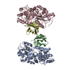 9fgvC  9fgyC  9fkqC M: map data used to model this data C: citing same article ( |
|---|---|
| Similar structure data |


-Links
 PDBj
PDBj


- Assembly
| Deposited unit | 
|
|---|---|
| 1 |
|
-Components
| #1: Antibody | Mass: 13863.432 Da / Num. of mol.: 2 Source method: isolated from a genetically manipulated source Source: (gene. exp.)  #2: Protein | Mass: 14331.160 Da / Num. of mol.: 2 / Source method: isolated from a natural source / Source: (natural) #3: Chemical |   Mass: 92.094 Da / Num. of mol.: 2 / Source method: obtained synthetically / Formula: C3H8O3 Mass: 92.094 Da / Num. of mol.: 2 / Source method: obtained synthetically / Formula: C3H8O3Has ligand of interest | N | Has protein modification | Y | |
|---|
-Experimental details
-Experiment
| Experiment | Method: ELECTRON MICROSCOPY |
|---|---|
| EM experiment | Aggregation state: PARTICLE / 3D reconstruction method: single particle reconstruction |
- Sample preparation
| Component | Name: Lysozyme homo-dimer assembled by homo Di-Gluebody GbMBP Type: COMPLEX / Entity ID: #1-#2 / Source: NATURAL | |||||||||||||||
|---|---|---|---|---|---|---|---|---|---|---|---|---|---|---|---|---|
| Molecular weight | Value: 0.057 MDa / Experimental value: NO | |||||||||||||||
| Source (natural) | Organism: | |||||||||||||||
| Source (recombinant) | Organism: | |||||||||||||||
| Buffer solution | pH: 7.5 | |||||||||||||||
| Buffer component |
| |||||||||||||||
| Specimen | Embedding applied: NO / Shadowing applied: NO / Staining applied: NO / Vitrification applied: YES | |||||||||||||||
| Specimen support | Grid type: Quantifoil R1.2/1.3 | |||||||||||||||
| Vitrification | Instrument: FEI VITROBOT MARK I / Cryogen name: ETHANE / Humidity: 100 % / Chamber temperature: 278 K |
- Electron microscopy imaging
| Experimental equipment |  Model: Titan Krios / Image courtesy: FEI Company |
|---|---|
| Microscopy | Model: FEI TITAN KRIOS |
| Electron gun | Electron source:  FIELD EMISSION GUN / Accelerating voltage: 300 kV / Illumination mode: FLOOD BEAM FIELD EMISSION GUN / Accelerating voltage: 300 kV / Illumination mode: FLOOD BEAM |
| Electron lens | Mode: BRIGHT FIELD / Nominal defocus max: 2600 nm / Nominal defocus min: 1600 nm / Cs: 2.7 mm |
| Image recording | Electron dose: 50 e/Å2 / Film or detector model: FEI FALCON IV (4k x 4k) |
| EM imaging optics | Energyfilter slit width: 20 eV |
- Processing
| EM software |
| |||||||||||||||||||||||||||
|---|---|---|---|---|---|---|---|---|---|---|---|---|---|---|---|---|---|---|---|---|---|---|---|---|---|---|---|---|
| CTF correction | Type: PHASE FLIPPING AND AMPLITUDE CORRECTION | |||||||||||||||||||||||||||
| 3D reconstruction | Resolution: 3.53 Å / Resolution method: FSC 0.143 CUT-OFF / Num. of particles: 269765 / Symmetry type: POINT | |||||||||||||||||||||||||||
| Atomic model building | Space: REAL | |||||||||||||||||||||||||||
| Atomic model building | PDB-ID: 6BJ2 Accession code: 6BJ2 / Source name: PDB / Type: experimental model | |||||||||||||||||||||||||||
| Refine LS restraints |
|

