 Movie
Movie Controller


+ Open data

- Basic information
| Entry | Database: PDB / ID: 8qv6 | |||||||||
|---|---|---|---|---|---|---|---|---|---|---|
| Title | Structure of human SPNS2 in DDM | |||||||||
 Components Components |
| |||||||||
 Keywords Keywords | LIPID TRANSPORT / SLC TRANSPORTER / MEMBRANE PROTEIN / S1P / EXPORTER | |||||||||
| Function / homology |  Function and homology information Function and homology informationregulation of eye pigmentation / regulation of humoral immune response / regulation of T cell migration / sphingolipid intramembrane carrier activity / lymphocyte migration / Sphingolipid de novo biosynthesis / sphingolipid biosynthetic process / sphingosine-1-phosphate receptor signaling pathway / lipid transport / B cell homeostasis ...regulation of eye pigmentation / regulation of humoral immune response / regulation of T cell migration / sphingolipid intramembrane carrier activity / lymphocyte migration / Sphingolipid de novo biosynthesis / sphingolipid biosynthetic process / sphingosine-1-phosphate receptor signaling pathway / lipid transport / B cell homeostasis / T cell homeostasis / lymph node development / transmembrane transporter activity / sensory perception of sound / bone development / endosome membrane / membrane / plasma membrane Similarity search - Function | |||||||||
| Biological species |  Homo sapiens (human) Homo sapiens (human) | |||||||||
| Method | ELECTRON MICROSCOPY / single particle reconstruction / cryo EM / Resolution: 3.68 Å | |||||||||
 Authors Authors | Li, H.Z. / Pike, A.C.W. / McKinley, G. / Mukhopadhyay, S.M.M. / Moreau, C. / Scacioc, A. / Abrusci, P. / Borkowska, O. / Chalk, R. / Stefanic, S. ...Li, H.Z. / Pike, A.C.W. / McKinley, G. / Mukhopadhyay, S.M.M. / Moreau, C. / Scacioc, A. / Abrusci, P. / Borkowska, O. / Chalk, R. / Stefanic, S. / Burgess-Brown, N. / Duerr, K.L. / Sauer, D.B. | |||||||||
| Funding support |  Switzerland, Switzerland,  United Kingdom, 2items United Kingdom, 2items
| |||||||||
 Citation Citation | Journal: Nat Commun / Year: 2025 Title: Transport and inhibition of the sphingosine-1-phosphate exporter SPNS2. Authors: Huanyu Z Li / Ashley C W Pike / Yung-Ning Chang / Dheeraj Prakaash / Zuzana Gelova / Josefina Stanka / Christophe Moreau / Hannah C Scott / Frank Wunder / Gernot Wolf / Andreea Scacioc / ...Authors: Huanyu Z Li / Ashley C W Pike / Yung-Ning Chang / Dheeraj Prakaash / Zuzana Gelova / Josefina Stanka / Christophe Moreau / Hannah C Scott / Frank Wunder / Gernot Wolf / Andreea Scacioc / Gavin McKinley / Helena Batoulis / Shubhashish Mukhopadhyay / Andrea Garofoli / Adán Pinto-Fernández / Benedikt M Kessler / Nicola A Burgess-Brown / Saša Štefanić / Tabea Wiedmer / Katharina L Dürr / Vera Puetter / Alexander Ehrmann / Syma Khalid / Alvaro Ingles-Prieto / Giulio Superti-Furga / David B Sauer /   Abstract: Sphingosine-1-phosphate (S1P) is a signaling lysolipid critical to heart development, immunity, and hearing. Accordingly, mutations in the S1P transporter SPNS2 are associated with reduced white cell ...Sphingosine-1-phosphate (S1P) is a signaling lysolipid critical to heart development, immunity, and hearing. Accordingly, mutations in the S1P transporter SPNS2 are associated with reduced white cell count and hearing defects. SPNS2 also exports the S1P-mimicking FTY720-P (Fingolimod) and thereby is central to the pharmacokinetics of this drug when treating multiple sclerosis. Here, we use a combination of cryo-electron microscopy, immunofluorescence, in vitro binding and in vivo S1P export assays, and molecular dynamics simulations to probe SPNS2's substrate binding and transport. These results reveal the transporter's binding mode to its native substrate S1P, the therapeutic FTY720-P, and the reported SPNS2-targeting inhibitor 33p. Further capturing an inward-facing apo state, our structures illuminate the protein's mechanism for exchange between inward-facing and outward-facing conformations. Finally, using these structural, localization, and S1P transport results, we identify how pathogenic mutations ablate the protein's export activity and thereby lead to hearing loss. | |||||||||
| History |
|
- Structure visualization
| Structure viewer | Molecule: MolmilJmol/JSmol |
|---|
- Downloads & links
-Download
| PDBx/mmCIF format | 8qv6.cif.gz | 127.5 KB | Display | PDBx/mmCIF format |
|---|---|---|---|---|
| PDB format | pdb8qv6.ent.gz | 78.4 KB | Display | PDB format |
| PDBx/mmJSON format | 8qv6.json.gz | Tree view | PDBx/mmJSON format | |
| Others |  Other downloads Other downloads |
-Validation report
| Arichive directory | https://data.pdbj.org/pub/pdb/validation_reports/qv/8qv6ftp://data.pdbj.org/pub/pdb/validation_reports/qv/8qv6 | HTTPS FTP |
|---|
-Related structure data
| Related structure data | 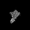 18668MC 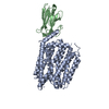 8qv5C M: map data used to model this data C: citing same article ( |
|---|---|
| Similar structure data |
-Links
 PDBj
PDBj


- Assembly
| Deposited unit | 
|
|---|---|
| 1 |
|
-Components
| #1: Protein | Mass: 58962.211 Da / Num. of mol.: 1 Source method: isolated from a genetically manipulated source Source: (gene. exp.) Homo sapiens (human) / Gene: SPNS2 / Plasmid: pHTBV1.1-CTGFP-SIII-10H / Cell (production host): Expi293F / Production host: Homo sapiens (human) / References: UniProt: Q8IVW8 |
|---|---|
| #2: Antibody | Mass: 15674.475 Da / Num. of mol.: 1 Source method: isolated from a genetically manipulated source Source: (gene. exp.)  |
| #3: Sugar | ChemComp-LMT /   Type: D-saccharide / Mass: 510.615 Da / Num. of mol.: 1 / Source method: obtained synthetically / Formula: C24H46O11 / Feature type: SUBJECT OF INVESTIGATION / Comment: detergent*YM Type: D-saccharide / Mass: 510.615 Da / Num. of mol.: 1 / Source method: obtained synthetically / Formula: C24H46O11 / Feature type: SUBJECT OF INVESTIGATION / Comment: detergent*YM |
| Has ligand of interest | Y |
| Has protein modification | Y |
-Experimental details
-Experiment
| Experiment | Method: ELECTRON MICROSCOPY |
|---|---|
| EM experiment | Aggregation state: PARTICLE / 3D reconstruction method: single particle reconstruction |
- Sample preparation
| Component | Name: Complex of SPNS2 with nanobody D12 / Type: COMPLEX / Entity ID: #1-#2 / Source: RECOMBINANT | ||||||||||||||||
|---|---|---|---|---|---|---|---|---|---|---|---|---|---|---|---|---|---|
| Molecular weight | Value: 0.07453 ° / Experimental value: NO | ||||||||||||||||
| Source (natural) | Organism: Homo sapiens (human) | ||||||||||||||||
| Source (recombinant) | Organism: Homo sapiens (human) | ||||||||||||||||
| Buffer solution | pH: 7.5 / Details: 20mM HEPES pH 7.5, 150mM NaCl, 0.025% DDM | ||||||||||||||||
| Buffer component |
| ||||||||||||||||
| Specimen | Conc.: 14 mg/ml / Embedding applied: NO / Shadowing applied: NO / Staining applied: NO / Vitrification applied: YES / Details: SEC purified | ||||||||||||||||
| Specimen support | Grid material: GOLD / Grid mesh size: 300 divisions/in. / Grid type: Quantifoil R1.2/1.3 | ||||||||||||||||
| Vitrification | Instrument: FEI VITROBOT MARK IV / Cryogen name: ETHANE / Humidity: 100 % / Chamber temperature: 277 K |
- Electron microscopy imaging
| Experimental equipment |  Model: Titan Krios / Image courtesy: FEI Company |
|---|---|
| Microscopy | Model: FEI TITAN KRIOS |
| Electron gun | Electron source:  FIELD EMISSION GUN / Accelerating voltage: 300 kV / Illumination mode: FLOOD BEAM FIELD EMISSION GUN / Accelerating voltage: 300 kV / Illumination mode: FLOOD BEAM |
| Electron lens | Mode: BRIGHT FIELD / Nominal magnification: 105000 X / Nominal defocus max: 2400 nm / Nominal defocus min: 1000 nm / C2 aperture diameter: 70 µm / Alignment procedure: COMA FREE |
| Specimen holder | Cryogen: NITROGEN / Specimen holder model: FEI TITAN KRIOS AUTOGRID HOLDER |
| Image recording | Average exposure time: 1.6 sec. / Electron dose: 25.4641 e/Å2 / Film or detector model: GATAN K3 (6k x 4k) / Num. of grids imaged: 1 / Num. of real images: 12014 / Details: eBIC Diamond Krios IV |
- Processing
| EM software |
| ||||||||||||||||||||||||||||||||||||||||||||||||
|---|---|---|---|---|---|---|---|---|---|---|---|---|---|---|---|---|---|---|---|---|---|---|---|---|---|---|---|---|---|---|---|---|---|---|---|---|---|---|---|---|---|---|---|---|---|---|---|---|---|
| CTF correction | Type: PHASE FLIPPING AND AMPLITUDE CORRECTION | ||||||||||||||||||||||||||||||||||||||||||||||||
| Particle selection | Num. of particles selected: 6672673 | ||||||||||||||||||||||||||||||||||||||||||||||||
| Symmetry | Point symmetry: C1 (asymmetric) | ||||||||||||||||||||||||||||||||||||||||||||||||
| 3D reconstruction | Resolution: 3.68 Å / Resolution method: FSC 0.143 CUT-OFF / Num. of particles: 246225 / Algorithm: FOURIER SPACE Details: Masked Local refinement in cryoSPARC with detergent micelle omitted Symmetry type: POINT | ||||||||||||||||||||||||||||||||||||||||||||||||
| Atomic model building | Protocol: OTHER / Space: REAL | ||||||||||||||||||||||||||||||||||||||||||||||||
| Atomic model building | Source name: AlphaFold / Type: in silico model | ||||||||||||||||||||||||||||||||||||||||||||||||
| Refinement | Cross valid method: NONE Stereochemistry target values: GeoStd + Monomer Library + CDL v1.2 | ||||||||||||||||||||||||||||||||||||||||||||||||
| Displacement parameters | Biso mean: 98.11 Å2 | ||||||||||||||||||||||||||||||||||||||||||||||||
| Refine LS restraints |
|
