ムービー
ムービー コントローラー


+ データを開く

- 基本情報
基本情報
| 登録情報 |  | |||||||||
|---|---|---|---|---|---|---|---|---|---|---|
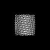
| タイトル | 400A Vipp1 H1-6 helical tubes | |||||||||
 マップデータ マップデータ | ||||||||||
 試料 試料 |
| |||||||||
 キーワード キーワード | membrane remodeling / membrane tubulation / LIPID BINDING PROTEIN | |||||||||
| 機能・相同性 | PspA/IM30 / PspA/IM30 family / plasma membrane / Membrane-associated protein Vipp1 機能・相同性情報 機能・相同性情報 | |||||||||
| 生物種 |  | |||||||||
| 手法 | らせん対称体再構成法 / クライオ電子顕微鏡法 / 解像度: 7.0 Å | |||||||||
 データ登録者 データ登録者 | Junglas B / Sachse C | |||||||||
| 資金援助 |  ドイツ, 2件 ドイツ, 2件
| |||||||||
 引用 引用 | ジャーナル: Nat Struct Mol Biol / 年: 2025 タイトル: Structural basis for Vipp1 membrane binding: from loose coats and carpets to ring and rod assemblies. 著者: Benedikt Junglas / David Kartte / Mirka Kutzner / Nadja Hellmann / Ilona Ritter / Dirk Schneider / Carsten Sachse / 要旨: Vesicle-inducing protein in plastids 1 (Vipp1) is critical for thylakoid membrane biogenesis and maintenance. Although Vipp1 has recently been identified as a member of the endosomal sorting ...Vesicle-inducing protein in plastids 1 (Vipp1) is critical for thylakoid membrane biogenesis and maintenance. Although Vipp1 has recently been identified as a member of the endosomal sorting complexes required for transport III superfamily, it is still unknown how Vipp1 remodels membranes. Here, we present cryo-electron microscopy structures of Synechocystis Vipp1 interacting with membranes: seven structures of helical and stacked-ring assemblies at 5-7-Å resolution engulfing membranes and three carpet structures covering lipid vesicles at ~20-Å resolution using subtomogram averaging. By analyzing ten structures of N-terminally truncated Vipp1, we show that helix α0 is essential for membrane tubulation and forms the membrane-anchoring domain of Vipp1. Lastly, using a conformation-restrained Vipp1 mutant, we reduced the structural plasticity of Vipp1 and determined two structures of Vipp1 at 3.0-Å resolution, resolving the molecular details of membrane-anchoring and intersubunit contacts of helix α0. Our data reveal membrane curvature-dependent structural transitions from carpets to rings and rods, some of which are capable of inducing and/or stabilizing high local membrane curvature triggering membrane fusion. #1: ジャーナル: Biorxiv / 年: 2024タイトル: Structural basis for Vipp1 membrane binding: From loose coats and carpets to ring and rod assemblies 著者: Junglas B / Kartte D / Kutzner M / Hellmann N / Ritter I / Schneider D / Sachse C | |||||||||
| 履歴 |
|
- 構造の表示
構造の表示
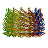
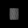
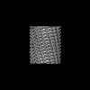
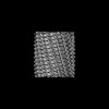
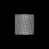
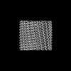
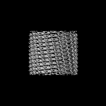
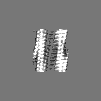
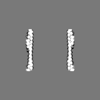
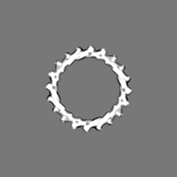
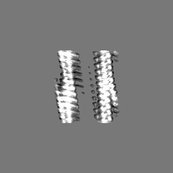
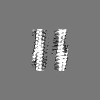
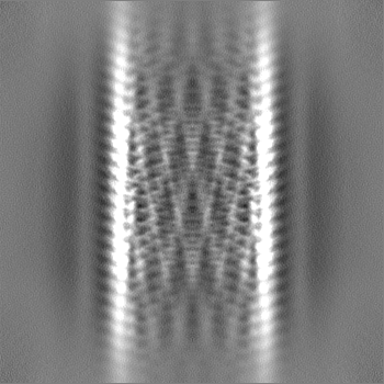
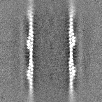
| 添付画像 |
|---|
- ダウンロードとリンク
-EMDBアーカイブ
| マップデータ | emd_18433.map.gz | 5.6 MB | EMDBマップデータ形式 | |
|---|---|---|---|---|
| ヘッダ (付随情報) | emd-18433-v30.xmlemd-18433.xml | 17.4 KB 17.4 KB | 表示 表示 | EMDBヘッダ |
| FSC (解像度算出) | emd_18433_fsc.xml | 11.6 KB | 表示 | FSCデータファイル |
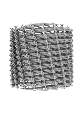
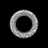
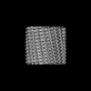
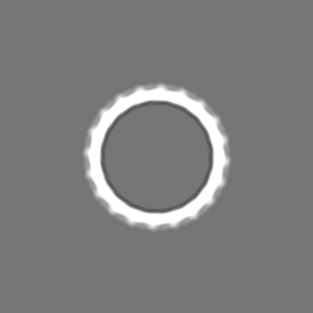
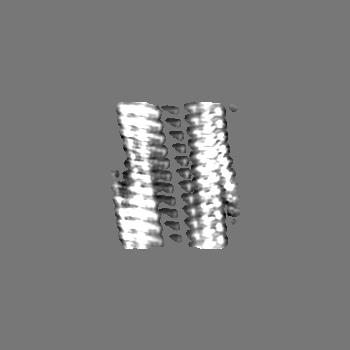
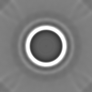
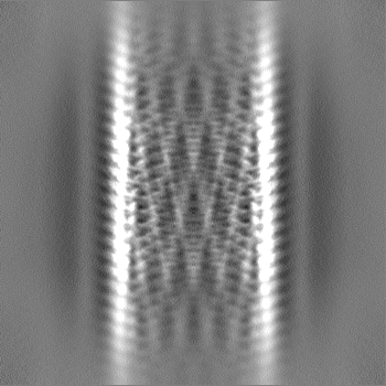
| 画像 |  emd_18433.png emd_18433.png | 69.2 KB | ||
| Filedesc metadata | emd-18433.cif.gz | 5.8 KB | ||
| その他 | emd_18433_half_map_1.map.gzemd_18433_half_map_2.map.gz | 151.7 MB 151.7 MB | ||
| アーカイブディレクトリ |  http://ftp.pdbj.org/pub/emdb/structures/EMD-18433ftp://ftp.pdbj.org/pub/emdb/structures/EMD-18433 http://ftp.pdbj.org/pub/emdb/structures/EMD-18433ftp://ftp.pdbj.org/pub/emdb/structures/EMD-18433 | HTTPS FTP |
-検証レポート
| 文書・要旨 | emd_18433_validation.pdf.gz | 812.4 KB | 表示 | EMDB検証レポート |
|---|---|---|---|---|
| 文書・詳細版 | emd_18433_full_validation.pdf.gz | 812 KB | 表示 | |
| XML形式データ | emd_18433_validation.xml.gz | 19.9 KB | 表示 | |
| CIF形式データ | emd_18433_validation.cif.gz | 26.1 KB | 表示 | |
| アーカイブディレクトリ | https://ftp.pdbj.org/pub/emdb/validation_reports/EMD-18433ftp://ftp.pdbj.org/pub/emdb/validation_reports/EMD-18433 | HTTPS FTP |
-関連構造データ
| 関連構造データ |  8qi4MC  8qfvC  8qhvC  8qhwC  8qhxC  8qhyC  8qhzC  8qi0C  8qi1C  8qi2C  8qi3C  8qi5C 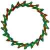 8qi6C  9eomC  9eonC  9eooC  9eopC M: このマップから作成された原子モデル C: 同じ文献を引用 ( |
|---|---|
| 類似構造データ |


-リンク
| EMDBのページ | EMDB (EBI/PDBe) / EMDataResource |
|---|
-マップ
| ファイル | ダウンロード / ファイル: emd_18433.map.gz / 形式: CCP4 / 大きさ: 163.6 MB / タイプ: IMAGE STORED AS FLOATING POINT NUMBER (4 BYTES) | ||||||||||||||||||||||||||||||||||||
|---|---|---|---|---|---|---|---|---|---|---|---|---|---|---|---|---|---|---|---|---|---|---|---|---|---|---|---|---|---|---|---|---|---|---|---|---|---|
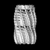
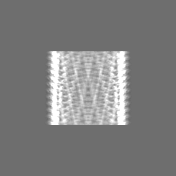
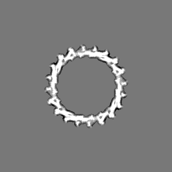
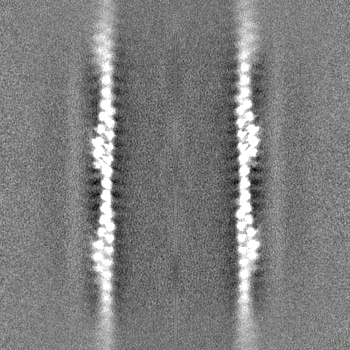
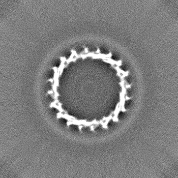
| 投影像・断面図 | 画像のコントロール
画像は Spider により作成 | ||||||||||||||||||||||||||||||||||||
| ボクセルのサイズ | X=Y=Z: 2.4 Å | ||||||||||||||||||||||||||||||||||||
| 密度 |
| ||||||||||||||||||||||||||||||||||||
| 対称性 | 空間群: 1 | ||||||||||||||||||||||||||||||||||||
| 詳細 | EMDB XML:
|
 Z (Sec.)
 Y (Row.)
 X (Col.)


-添付データ
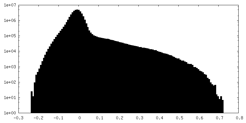
-ハーフマップ: #2
| ファイル | emd_18433_half_map_1.map | ||||||||||||
|---|---|---|---|---|---|---|---|---|---|---|---|---|---|
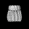
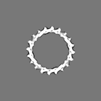
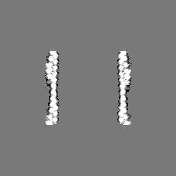
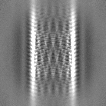
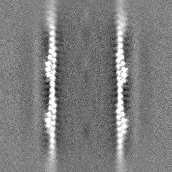
| 投影像・断面図 |
| ||||||||||||
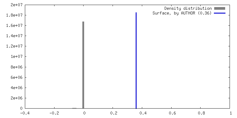
| 密度ヒストグラム |


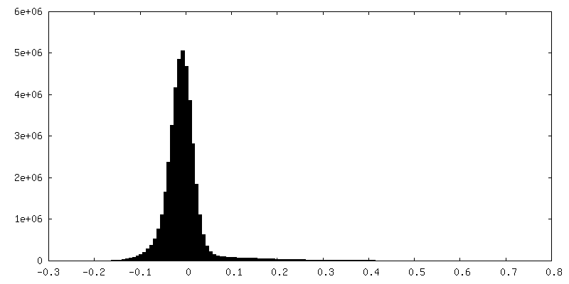
-ハーフマップ: #1
| ファイル | emd_18433_half_map_2.map | ||||||||||||
|---|---|---|---|---|---|---|---|---|---|---|---|---|---|
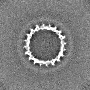
| 投影像・断面図 |
| ||||||||||||
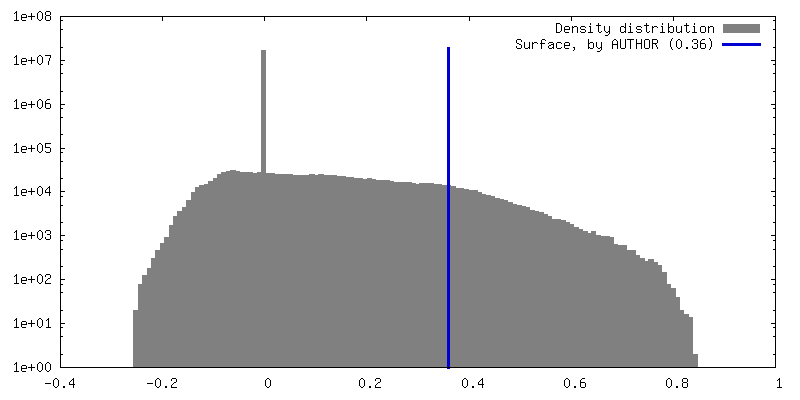
| 密度ヒストグラム |


- 試料の構成要素
-全体 : Vipp1 H1-6
| 全体 | 名称: Vipp1 H1-6 |
|---|---|
| 要素 |
|
-超分子 #1: Vipp1 H1-6
| 超分子 | 名称: Vipp1 H1-6 / タイプ: complex / ID: 1 / 親要素: 0 / 含まれる分子: all |
|---|---|
| 由来(天然) | 生物種: |
-分子 #1: Membrane-associated protein Vipp1
| 分子 | 名称: Membrane-associated protein Vipp1 / タイプ: protein_or_peptide / ID: 1 / コピー数: 1 / 光学異性体: LEVO |
|---|---|
| 由来(天然) | 生物種: |
| 分子量 | 理論値: 21.060562 KDa |
| 組換発現 | 生物種: |
| 配列 | 文字列: DPEKVLEQAV IDMQEDLVQL RQAVARTIAE EKRTEQRLNQ DTQEAKKWED RAKLALTNGE ENLAREALAR KKSLTDTAAA YQTQLAQQR TMSENLRRNL AALEAKISEA KTKKNMLQAR AKAAKANAEL QQTLGGLGTS SATSAFERME NKVLDMEATS Q AAGELAGF ...文字列: DPEKVLEQAV IDMQEDLVQL RQAVARTIAE EKRTEQRLNQ DTQEAKKWED RAKLALTNGE ENLAREALAR KKSLTDTAAA YQTQLAQQR TMSENLRRNL AALEAKISEA KTKKNMLQAR AKAAKANAEL QQTLGGLGTS SATSAFERME NKVLDMEATS Q AAGELAGF GIENQFAQLE ASSGVEDELA ALKA UniProtKB: Membrane-associated protein Vipp1 |
-実験情報
-構造解析
| 手法 | クライオ電子顕微鏡法 |
|---|---|
 解析 解析 | らせん対称体再構成法 |
| 試料の集合状態 | helical array |
-試料調製
| 緩衝液 | pH: 8 |
|---|---|
| 糖包埋 | 材質: vitreous ice |
| 凍結 | 凍結剤: ETHANE |
- 電子顕微鏡法
| 顕微鏡 | FEI TITAN KRIOS |
|---|---|
| 撮影 | フィルム・検出器のモデル: FEI FALCON IV (4k x 4k) 平均電子線量: 48.0 e/Å2 |
| 電子線 | 加速電圧: 300 kV / 電子線源:  FIELD EMISSION GUN FIELD EMISSION GUN |
| 電子光学系 | 照射モード: FLOOD BEAM / 撮影モード: BRIGHT FIELD / 最大 デフォーカス(公称値): 2.0 µm / 最小 デフォーカス(公称値): 1.0 µm |
| 実験機器 |  モデル: Titan Krios / 画像提供: FEI Company |
-画像解析
| 最終 再構成 | 想定した対称性 - らせんパラメータ - Δz: 6.03 Å 想定した対称性 - らせんパラメータ - ΔΦ: -17.6 ° 想定した対称性 - らせんパラメータ - 軸対称性: C4 (4回回転対称) 解像度のタイプ: BY AUTHOR / 解像度: 7.0 Å / 解像度の算出法: FSC 0.143 CUT-OFF / 使用した粒子像数: 77818 |
|---|---|
| 初期モデル | モデルのタイプ: PDB ENTRY PDBモデル - PDB ID: |
| 最終 角度割当 | タイプ: NOT APPLICABLE |
| FSC曲線 (解像度の算出) |  |