 ムービー
ムービー コントローラー


+ データを開く

- 基本情報
基本情報
| 登録情報 | 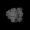 | |||||||||
|---|---|---|---|---|---|---|---|---|---|---|
| タイトル | 70S ribosome with A, P and E-site tRNAs in chloramphenicol-treated Mycoplasma pneumoniae cells, K3 data | |||||||||
 マップデータ マップデータ | ||||||||||
 試料 試料 |
| |||||||||
 キーワード キーワード | In situ / cryo-electron tomography / bacterial ribosome / Chloramphenicol / antibiotic / RIBOSOME | |||||||||
| 生物種 |  Mycoplasmoides pneumoniae M129 (バクテリア) Mycoplasmoides pneumoniae M129 (バクテリア) | |||||||||
| 手法 | サブトモグラム平均法 / クライオ電子顕微鏡法 / 解像度: 3.8 Å | |||||||||
 データ登録者 データ登録者 | Xue L / Spahn C / Schacherl M / Mahamid J | |||||||||
| 資金援助 |  米国, 米国,  ドイツ, 2件 ドイツ, 2件
| |||||||||
 引用 引用 | ジャーナル: Nat Struct Mol Biol / 年: 2025 タイトル: Structural insights into context-dependent inhibitory mechanisms of chloramphenicol in cells. 著者: Liang Xue / Christian M T Spahn / Magdalena Schacherl / Julia Mahamid /  要旨: Ribosome-targeting antibiotics represent an important class of antimicrobial drugs. Chloramphenicol (Cm) is a well-studied ribosomal peptidyl transferase center (PTC) binder and growing evidence ...Ribosome-targeting antibiotics represent an important class of antimicrobial drugs. Chloramphenicol (Cm) is a well-studied ribosomal peptidyl transferase center (PTC) binder and growing evidence suggests that its inhibitory action depends on the sequence of the nascent peptide. How such selective inhibition on the molecular scale manifests on the cellular level remains unclear. Here, we use cryo-electron tomography to analyze the impact of Cm inside the bacterium Mycoplasma pneumoniae. By resolving the Cm-bound ribosomes to 3.0 Å, we elucidate Cm's coordination with natural nascent peptides and transfer RNAs in the PTC. We find that Cm leads to the accumulation of a number of translation elongation states, indicating ongoing futile accommodation cycles, and to extensive ribosome collisions. We, thus, suggest that, beyond its direct inhibition of protein synthesis, the action of Cm may involve the activation of cellular stress responses. This work exemplifies how in-cell structural biology can expand the understanding of mechanisms of action for extensively studied antibiotics. | |||||||||
| 履歴 |
|
- 構造の表示
構造の表示
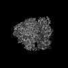
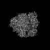
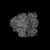
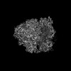
| 添付画像 |
|---|

- ダウンロードとリンク
-EMDBアーカイブ
| マップデータ | emd_17137.map.gz | 7.6 MB |  EMDBマップデータ形式 EMDBマップデータ形式 | |
|---|---|---|---|---|
| ヘッダ (付随情報) | emd-17137-v30.xmlemd-17137.xml | 19.5 KB 19.5 KB | 表示 表示 | EMDBヘッダ |
| FSC (解像度算出) | emd_17137_fsc.xml | 9.1 KB | 表示 | FSCデータファイル |
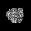
| 画像 |  emd_17137.png emd_17137.png | 225.3 KB | ||
| マスクデータ | emd_17137_msk_1.map | 64 MB | マスクマップ | |
| Filedesc metadata | emd-17137.cif.gz | 5.3 KB | ||
| その他 | emd_17137_half_map_1.map.gzemd_17137_half_map_2.map.gz | 49.6 MB 49.6 MB | ||
| アーカイブディレクトリ |  http://ftp.pdbj.org/pub/emdb/structures/EMD-17137ftp://ftp.pdbj.org/pub/emdb/structures/EMD-17137 http://ftp.pdbj.org/pub/emdb/structures/EMD-17137ftp://ftp.pdbj.org/pub/emdb/structures/EMD-17137 | HTTPS FTP |
-検証レポート
| 文書・要旨 | emd_17137_validation.pdf.gz | 165.3 KB | 表示 | EMDB検証レポート |
|---|---|---|---|---|
| 文書・詳細版 | emd_17137_full_validation.pdf.gz | 164.9 KB | 表示 | |
| XML形式データ | emd_17137_validation.xml.gz | 571 B | 表示 | |
| CIF形式データ | emd_17137_validation.cif.gz | 483 B | 表示 | |
| アーカイブディレクトリ | https://ftp.pdbj.org/pub/emdb/validation_reports/EMD-17137ftp://ftp.pdbj.org/pub/emdb/validation_reports/EMD-17137 | HTTPS FTP |
-関連構造データ


-リンク
| EMDBのページ | EMDB (EBI/PDBe) / EMDataResource |
|---|
-マップ
| ファイル | ダウンロード / ファイル: emd_17137.map.gz / 形式: CCP4 / 大きさ: 64 MB / タイプ: IMAGE STORED AS FLOATING POINT NUMBER (4 BYTES) | ||||||||||||||||||||||||||||||||||||
|---|---|---|---|---|---|---|---|---|---|---|---|---|---|---|---|---|---|---|---|---|---|---|---|---|---|---|---|---|---|---|---|---|---|---|---|---|---|
| 投影像・断面図 | 画像のコントロール
画像は Spider により作成 | ||||||||||||||||||||||||||||||||||||
| ボクセルのサイズ | X=Y=Z: 1.7005 Å | ||||||||||||||||||||||||||||||||||||
| 密度 |
| ||||||||||||||||||||||||||||||||||||
| 対称性 | 空間群: 1 | ||||||||||||||||||||||||||||||||||||
| 詳細 | EMDB XML:
|
 Z (Sec.)
 Y (Row.)
 X (Col.)


-添付データ
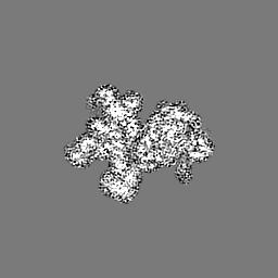
-マスク #1
| ファイル | emd_17137_msk_1.map | ||||||||||||
|---|---|---|---|---|---|---|---|---|---|---|---|---|---|
| 投影像・断面図 |
| ||||||||||||
| 密度ヒストグラム |


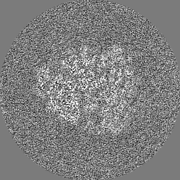
-ハーフマップ: #1
| ファイル | emd_17137_half_map_1.map | ||||||||||||
|---|---|---|---|---|---|---|---|---|---|---|---|---|---|
| 投影像・断面図 |
| ||||||||||||
| 密度ヒストグラム |


-ハーフマップ: #2
| ファイル | emd_17137_half_map_2.map | ||||||||||||
|---|---|---|---|---|---|---|---|---|---|---|---|---|---|
| 投影像・断面図 |
| ||||||||||||
| 密度ヒストグラム |


- 試料の構成要素
-全体 : Mycoplasma pneumoniae M129 cells treated with chloramphenicol
| 全体 | 名称: Mycoplasma pneumoniae M129 cells treated with chloramphenicol |
|---|---|
| 要素 |
|
-超分子 #1: Mycoplasma pneumoniae M129 cells treated with chloramphenicol
| 超分子 | 名称: Mycoplasma pneumoniae M129 cells treated with chloramphenicol タイプ: cell / ID: 1 / 親要素: 0 |
|---|---|
| 由来(天然) | 生物種: Mycoplasmoides pneumoniae M129 (バクテリア) |
-実験情報
-構造解析
| 手法 | クライオ電子顕微鏡法 |
|---|---|
 解析 解析 | サブトモグラム平均法 |
| 試料の集合状態 | cell |
-試料調製
| 緩衝液 | pH: 7.4 詳細: Modified Hayflick medium: 14.7g/l Difco PPLO(Becton Dickinson), 20% (v/v) Gibco horse serum (New Zealand origin), 100 mM HEPES-Na; pH 7.4, 1% (w/w) glucose, 0.002% (w/w) phenol red, 1000 U/ml penicillin G. |
|---|---|
| グリッド | モデル: Quantifoil R2/1 / 材質: GOLD / メッシュ: 200 / 支持フィルム - 材質: CARBON / 支持フィルム - トポロジー: HOLEY |
| 凍結 | 凍結剤: ETHANE-PROPANE / 装置: HOMEMADE PLUNGER 詳細: Back-side blotting for 2-3 seconds before plunging using a manual plunger without an environmental control chamber.. |
| 詳細 | Mycoplasma pneumoniae M129 cells were grown on gold Quantifoil grids at 37 Celsius in modified Hayflick medium. Treatment with chloramphenicol at a final concentration of 0.2 mg/ml was performed for approximately 15 minutes before plunge freezing. |
- 電子顕微鏡法
| 顕微鏡 | FEI TITAN KRIOS |
|---|---|
| 特殊光学系 | エネルギーフィルター - 名称: GIF Bioquantum / エネルギーフィルター - スリット幅: 20 eV |
| 撮影 | フィルム・検出器のモデル: GATAN K3 BIOQUANTUM (6k x 4k) 実像数: 1 / 平均電子線量: 3.34 e/Å2 詳細: Gatan K3 camera in non-CDS counting mode, targeted dose rate on camera ~20 e/pixel/second, 10 frames per tilt image, constant exposure time for each tilt, pixel size 1.329A |
| 電子線 | 加速電圧: 300 kV / 電子線源:  FIELD EMISSION GUN FIELD EMISSION GUN |
| 電子光学系 | 照射モード: FLOOD BEAM / 撮影モード: BRIGHT FIELD / Cs: 2.7 mm / 最大 デフォーカス(公称値): 3.25 µm / 最小 デフォーカス(公称値): 1.0 µm / 倍率(公称値): 64000 |
| 試料ステージ | 試料ホルダーモデル: FEI TITAN KRIOS AUTOGRID HOLDER ホルダー冷却材: NITROGEN |
| 実験機器 |  モデル: Titan Krios / 画像提供: FEI Company |

