Movie
Movie Controller

[English] 日本語
 Yorodumi
- EMDB-1676: CryoEM 3D reconstruction of Rhodobacter capsulatus Mg-chelatase B... -
+ Open data

- Basic information
| Entry | Database: EMDB / ID: EMD-1676 | |||||||||
|---|---|---|---|---|---|---|---|---|---|---|
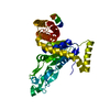
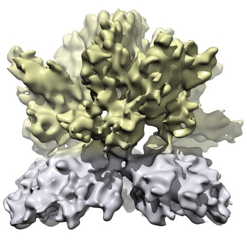
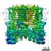
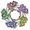
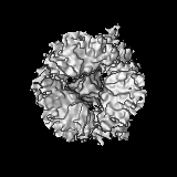
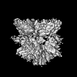
| Title | CryoEM 3D reconstruction of Rhodobacter capsulatus Mg-chelatase BchID complex in the presence of ADP | |||||||||
 Map data Map data | This is the volume of the complex of Magnesium Chelatase subunit BchI and BchD incubated with ADP. | |||||||||
 Sample Sample |
| |||||||||
 Keywords Keywords | AAA+ atpase / metallation / tetrapyrroles / Mg chelatase | |||||||||
| Function / homology |  Function and homology information Function and homology informationbacteriochlorophyll biosynthetic process / magnesium chelatase / magnesium chelatase activity / photosynthesis / ATP binding Similarity search - Function | |||||||||
| Biological species |  Rhodobacter capsulatus (bacteria) Rhodobacter capsulatus (bacteria) | |||||||||
| Method | single particle reconstruction / cryo EM / negative staining / Resolution: 7.5 Å | |||||||||
 Authors Authors | Lundqvist J / Elmlund H / Peterson-Wulff R / Elmlund D / Emanuelsson C / Hebert H / Willows R / Hansson M / Lindahl M / Al-Karadaghi S | |||||||||
 Citation Citation | Journal: J Mol Biol / Year: 2008 Title: A new cryo-EM single-particle ab initio reconstruction method visualizes secondary structure elements in an ATP-fueled AAA+ motor. Authors: Hans Elmlund / Joakim Lundqvist / Salam Al-Karadaghi / Mats Hansson / Hans Hebert / Martin Lindahl /  Abstract: The generation of ab initio three-dimensional (3D) models is a bottleneck in the studies of large macromolecular assemblies by single-particle cryo-electron microscopy. We describe here a novel ...The generation of ab initio three-dimensional (3D) models is a bottleneck in the studies of large macromolecular assemblies by single-particle cryo-electron microscopy. We describe here a novel method, in which established methods for two-dimensional image processing are combined with newly developed programs for joint rotational 3D alignment of a large number of class averages (RAD) and calculation of 3D volumes from aligned projections (VolRec). We demonstrate the power of the method by reconstructing an approximately 660-kDa ATP-fueled AAA+ motor to 7.5 A resolution, with secondary structure elements identified throughout the structure. We propose the method as a generally applicable automated strategy to obtain 3D reconstructions from unstained single particles imaged in vitreous ice. | |||||||||
| History |
|
- Structure visualization
| Movie |
Movie viewer |
|---|---|
| Structure viewer | EM map: SurfViewMolmilJmol/JSmol |
| Supplemental images |
- Downloads & links
-EMDB archive
| Map data | emd_1676.map.gz | 14 MB | EMDB map data format | |
|---|---|---|---|---|
| Header (meta data) | emd-1676-v30.xmlemd-1676.xml | 10 KB 10 KB | Display Display | EMDB header |
| Images |  emd_1676.jpg emd_1676.jpg | 47.3 KB | ||
| Archive directory |  http://ftp.pdbj.org/pub/emdb/structures/EMD-1676ftp://ftp.pdbj.org/pub/emdb/structures/EMD-1676 http://ftp.pdbj.org/pub/emdb/structures/EMD-1676ftp://ftp.pdbj.org/pub/emdb/structures/EMD-1676 | HTTPS FTP |
-Related structure data
| Related structure data | 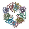 2x31MC 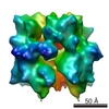 1677C  1678C C: citing same article ( M: atomic model generated by this map |
|---|---|
| Similar structure data |


-Links
| EMDB pages | EMDB (EBI/PDBe) / EMDataResource |
|---|---|
| Related items in Molecule of the Month |


-Map
| File | Download / File: emd_1676.map.gz / Format: CCP4 / Size: 15.3 MB / Type: IMAGE STORED AS FLOATING POINT NUMBER (4 BYTES) | ||||||||||||||||||||||||||||||||||||||||||||||||||||||||||||||||||||
|---|---|---|---|---|---|---|---|---|---|---|---|---|---|---|---|---|---|---|---|---|---|---|---|---|---|---|---|---|---|---|---|---|---|---|---|---|---|---|---|---|---|---|---|---|---|---|---|---|---|---|---|---|---|---|---|---|---|---|---|---|---|---|---|---|---|---|---|---|---|
| Annotation | This is the volume of the complex of Magnesium Chelatase subunit BchI and BchD incubated with ADP. | ||||||||||||||||||||||||||||||||||||||||||||||||||||||||||||||||||||
| Projections & slices | Image control
Images are generated by Spider. | ||||||||||||||||||||||||||||||||||||||||||||||||||||||||||||||||||||
| Voxel size | X=Y=Z: 1.167 Å | ||||||||||||||||||||||||||||||||||||||||||||||||||||||||||||||||||||
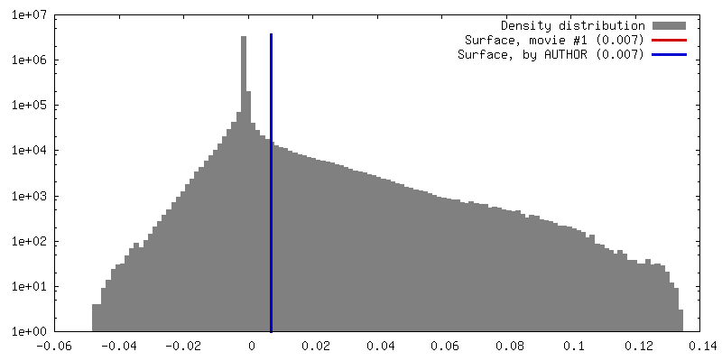
| Density |
| ||||||||||||||||||||||||||||||||||||||||||||||||||||||||||||||||||||
| Symmetry | Space group: 1 | ||||||||||||||||||||||||||||||||||||||||||||||||||||||||||||||||||||
| Details | EMDB XML:
CCP4 map header:
| ||||||||||||||||||||||||||||||||||||||||||||||||||||||||||||||||||||
 Z (Sec.)
 Y (Row.)
 X (Col.)


-Supplemental data
- Sample components
-Entire : Complex of Mg-chelatase subunits BchI and BchD in presence of ADP
| Entire | Name: Complex of Mg-chelatase subunits BchI and BchD in presence of ADP |
|---|---|
| Components |
|
-Supramolecule #1000: Complex of Mg-chelatase subunits BchI and BchD in presence of ADP
| Supramolecule | Name: Complex of Mg-chelatase subunits BchI and BchD in presence of ADP type: sample / ID: 1000 / Number unique components: 1 |
|---|---|
| Molecular weight | Experimental: 660 KDa / Theoretical: 660 KDa |
-Macromolecule #1: Biosynthetic enzyme
| Macromolecule | Name: Biosynthetic enzyme / type: protein_or_peptide / ID: 1 / Name.synonym: Mg chelatase / Recombinant expression: Yes |
|---|---|
| Source (natural) | Organism: Rhodobacter capsulatus (bacteria) |
| Molecular weight | Theoretical: 660 KDa |
| Recombinant expression | Organism: |
-Experimental details
-Structure determination
| Method | negative staining, cryo EM |
|---|---|
 Processing Processing | single particle reconstruction |
| Aggregation state | particle |
-Sample preparation
| Concentration | 0.1 mg/mL |
|---|---|
| Buffer | pH: 8 |
| Staining | Type: NEGATIVE / Details: Vitrification |
| Vitrification | Cryogen name: ETHANE / Instrument: OTHER |
- Electron microscopy
| Microscope | JEOL 2010F |
|---|---|
| Image recording | Digitization - Scanner: ZEISS SCAI / Number real images: 18 |
| Electron beam | Acceleration voltage: 120 kV / Electron source:  FIELD EMISSION GUN FIELD EMISSION GUN |
| Electron optics | Illumination mode: OTHER / Imaging mode: BRIGHT FIELD / Nominal defocus max: 5.5 µm / Nominal defocus min: 1.0 µm / Nominal magnification: 60000 |
| Sample stage | Specimen holder: Eucentric / Specimen holder model: JEOL |
-Image processing
| Final reconstruction | Applied symmetry - Point group: C3 (3 fold cyclic) / Resolution.type: BY AUTHOR / Resolution: 7.5 Å / Resolution method: FSC 0.5 CUT-OFF / Number images used: 29400 |
|---|---|
| Final two d classification | Number classes: 258 |