 ムービー
ムービー コントローラー


+ データを開く

- 基本情報
基本情報
| 登録情報 |  | |||||||||
|---|---|---|---|---|---|---|---|---|---|---|
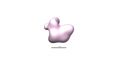
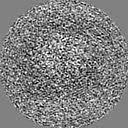
| タイトル | In situ subtomogram average of floating microtubule inner protein 16 (fMIP16) | |||||||||
 マップデータ マップデータ | In situ subtomogram average of floating microtubule inner protein 16 | |||||||||
 試料 試料 |
| |||||||||
 キーワード キーワード | microtubule associated protein / STRUCTURAL PROTEIN | |||||||||
| 生物種 |  Homo sapiens (ヒト) Homo sapiens (ヒト) | |||||||||
| 手法 | サブトモグラム平均法 / クライオ電子顕微鏡法 / 解像度: 27.0 Å | |||||||||
 データ登録者 データ登録者 | Chakraborty S / Mahamid J / Martinez-Sanchez A / Baumeister W | |||||||||
| 資金援助 | 1件
| |||||||||
 引用 引用 | ジャーナル: Proc Natl Acad Sci U S A / 年: 2025 タイトル: Cryo-ET suggests tubulin chaperones form a subset of microtubule lumenal particles with a role in maintaining neuronal microtubules. 著者: Saikat Chakraborty / Antonio Martinez-Sanchez / Florian Beck / Mauricio Toro-Nahuelpan / In-Young Hwang / Kyung-Min Noh / Wolfgang Baumeister / Julia Mahamid /  要旨: The functional architecture of the long-lived neuronal microtubule (MT) cytoskeleton is maintained by various MT-associated proteins (MAPs), most of which are known to bind to the MT outer surface. ...The functional architecture of the long-lived neuronal microtubule (MT) cytoskeleton is maintained by various MT-associated proteins (MAPs), most of which are known to bind to the MT outer surface. However, electron microscopy (EM) has long ago revealed the presence of particles inside the lumens of neuronal MTs, of yet unknown identity and function. Here, we use cryogenic electron tomography (cryo-ET) to analyze the three-dimensional (3D) organization and structures of MT lumenal particles in primary hippocampal neurons, human induced pluripotent stem cell-derived neurons, and pluripotent and differentiated P19 cells. We obtain in situ density maps of several lumenal particles from the respective cells and detect common structural features underscoring their potential overarching functions. Mass spectrometry-based proteomics combined with structural modeling suggest that a subset of lumenal particles could be tubulin-binding cofactors (TBCs) bound to tubulin monomers. A different subset of smaller particles, which remains unidentified, exhibits densities that bridge across the MT protofilaments. We show that increased lumenal particle concentration within MTs is concomitant with neuronal differentiation and correlates with higher MT curvatures. Enrichment of lumenal particles around MT lattice defects and at freshly polymerized MT open-ends suggests a MT protective role. Together with the identified structural resemblance of a subset of particles to TBCs, these results hint at a role in local tubulin proteostasis for the maintenance of long-lived neuronal MTs. #1: ジャーナル: Biorxiv / 年: 2024タイトル: Cryo-electron tomography suggests tubulin chaperones form a subset of microtubule lumenal particles with a role in maintaining neuronal microtubules 著者: Chakraborty S / Martinez-Sanchez A / Beck F / Toro-Nahuelpan M / Hwang IY / Noh KM / Baumeister W / Mahamid J | |||||||||
| 履歴 |
|
- 構造の表示
構造の表示
| 添付画像 |
|---|
- ダウンロードとリンク
-EMDBアーカイブ
| マップデータ | emd_15468.map.gz | 2.1 MB |  EMDBマップデータ形式 EMDBマップデータ形式 | |
|---|---|---|---|---|
| ヘッダ (付随情報) | emd-15468-v30.xmlemd-15468.xml | 16.6 KB 16.6 KB | 表示 表示 | EMDBヘッダ |
| FSC (解像度算出) | emd_15468_fsc.xml | 4.7 KB | 表示 | FSCデータファイル |
| 画像 |  emd_15468.png emd_15468.png | 9.3 KB | ||
| Filedesc metadata | emd-15468.cif.gz | 4.3 KB | ||
| その他 | emd_15468_half_map_1.map.gzemd_15468_half_map_2.map.gz | 6 MB 6 MB | ||
| アーカイブディレクトリ |  http://ftp.pdbj.org/pub/emdb/structures/EMD-15468ftp://ftp.pdbj.org/pub/emdb/structures/EMD-15468 http://ftp.pdbj.org/pub/emdb/structures/EMD-15468ftp://ftp.pdbj.org/pub/emdb/structures/EMD-15468 | HTTPS FTP |
-検証レポート
| 文書・要旨 | emd_15468_validation.pdf.gz | 835.8 KB | 表示 | EMDB検証レポート |
|---|---|---|---|---|
| 文書・詳細版 | emd_15468_full_validation.pdf.gz | 835.4 KB | 表示 | |
| XML形式データ | emd_15468_validation.xml.gz | 10.5 KB | 表示 | |
| CIF形式データ | emd_15468_validation.cif.gz | 14 KB | 表示 | |
| アーカイブディレクトリ | https://ftp.pdbj.org/pub/emdb/validation_reports/EMD-15468ftp://ftp.pdbj.org/pub/emdb/validation_reports/EMD-15468 | HTTPS FTP |
-関連構造データ
| 関連構造データ | C: 同じ文献を引用 ( |
|---|


-リンク
| EMDBのページ | EMDB (EBI/PDBe) / EMDataResource |
|---|
-マップ
| ファイル | ダウンロード / ファイル: emd_15468.map.gz / 形式: CCP4 / 大きさ: 8 MB / タイプ: IMAGE STORED AS FLOATING POINT NUMBER (4 BYTES) | ||||||||||||||||||||||||||||||||||||
|---|---|---|---|---|---|---|---|---|---|---|---|---|---|---|---|---|---|---|---|---|---|---|---|---|---|---|---|---|---|---|---|---|---|---|---|---|---|
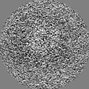
| 注釈 | In situ subtomogram average of floating microtubule inner protein 16 | ||||||||||||||||||||||||||||||||||||
| 投影像・断面図 | 画像のコントロール
画像は Spider により作成 | ||||||||||||||||||||||||||||||||||||
| ボクセルのサイズ | X=Y=Z: 2.129 Å | ||||||||||||||||||||||||||||||||||||
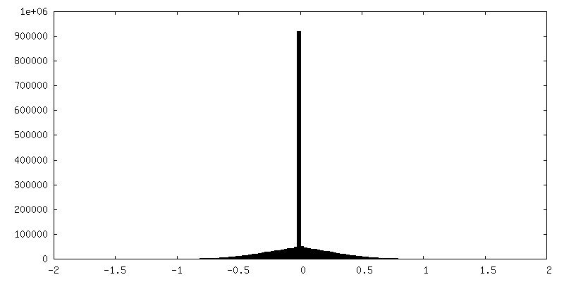
| 密度 |
| ||||||||||||||||||||||||||||||||||||
| 対称性 | 空間群: 1 | ||||||||||||||||||||||||||||||||||||
| 詳細 | EMDB XML:
|
 Z (Sec.)
 Y (Row.)
 X (Col.)


-添付データ
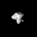
-ハーフマップ: In situ subtomogram average of floating microtubule inner protein 16
| ファイル | emd_15468_half_map_1.map | ||||||||||||
|---|---|---|---|---|---|---|---|---|---|---|---|---|---|
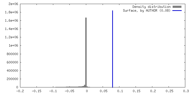
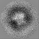
| 注釈 | In situ subtomogram average of floating microtubule inner protein 16 | ||||||||||||
| 投影像・断面図 |
| ||||||||||||
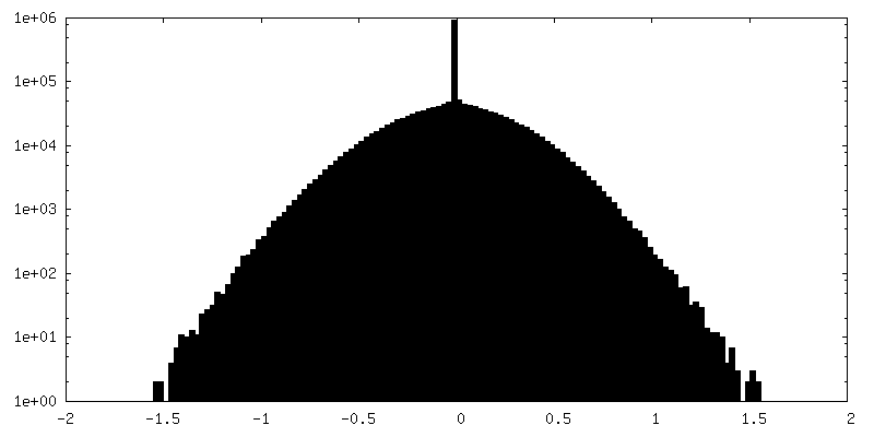
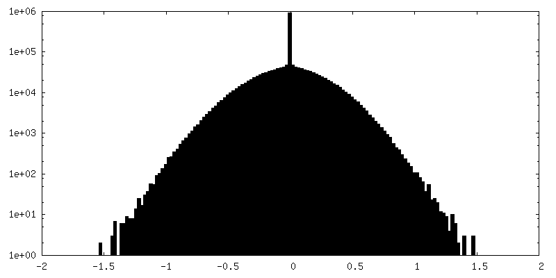
| 密度ヒストグラム |
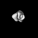
-ハーフマップ: In situ subtomogram average of floating microtubule inner protein 16
| ファイル | emd_15468_half_map_2.map | ||||||||||||
|---|---|---|---|---|---|---|---|---|---|---|---|---|---|
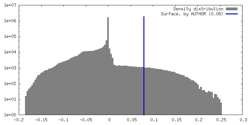
| 注釈 | In situ subtomogram average of floating microtubule inner protein 16 | ||||||||||||
| 投影像・断面図 |
| ||||||||||||
| 密度ヒストグラム |

- 試料の構成要素
-全体 : fMIP16 from human induced pluripotent stem cells derived neuronal...
| 全体 | 名称: fMIP16 from human induced pluripotent stem cells derived neuronal microtubules |
|---|---|
| 要素 |
|
-超分子 #1: fMIP16 from human induced pluripotent stem cells derived neuronal...
| 超分子 | 名称: fMIP16 from human induced pluripotent stem cells derived neuronal microtubules タイプ: cell / ID: 1 / 親要素: 0 |
|---|---|
| 由来(天然) | 生物種: Homo sapiens (ヒト) |
-実験情報
-構造解析
| 手法 | クライオ電子顕微鏡法 |
|---|---|
 解析 解析 | サブトモグラム平均法 |
| 試料の集合状態 | cell |
-試料調製
| 緩衝液 | pH: 7.2 |
|---|---|
| グリッド | モデル: Quantifoil / 材質: GOLD / メッシュ: 200 / 前処理 - タイプ: GLOW DISCHARGE / 前処理 - 時間: 30 sec. |
| 凍結 | 凍結剤: ETHANE-PROPANE / チャンバー内湿度: 70 % / チャンバー内温度: 310 K / 装置: LEICA EM GP |
| 詳細 | Frozen hydrated human induced pluripotent stem cells derived neurons |
- 電子顕微鏡法
| 顕微鏡 | FEI TITAN KRIOS |
|---|---|
| 温度 | 最低: 77.0 K |
| 特殊光学系 | 位相板: VOLTA PHASE PLATE / エネルギーフィルター - 名称: GIF Quantum LS / エネルギーフィルター - スリット幅: 20 eV |
| 撮影 | フィルム・検出器のモデル: GATAN K2 SUMMIT (4k x 4k) 検出モード: COUNTING / 平均電子線量: 2.0 e/Å2 |
| 電子線 | 加速電圧: 300 kV / 電子線源:  FIELD EMISSION GUN FIELD EMISSION GUN |
| 電子光学系 | C2レンズ絞り径: 70.0 µm / 照射モード: FLOOD BEAM / 撮影モード: BRIGHT FIELD / Cs: 2.7 mm / 最大 デフォーカス(公称値): 4.0 µm / 最小 デフォーカス(公称値): 2.5 µm / 倍率(公称値): 64000 |
| 試料ステージ | 試料ホルダーモデル: FEI TITAN KRIOS AUTOGRID HOLDER ホルダー冷却材: NITROGEN |
| 実験機器 |  モデル: Titan Krios / 画像提供: FEI Company |

