+ データを開く
- 基本情報
基本情報
| 登録情報 | データベース: EMDB / ID: EMD-1545 | |||||||||
|---|---|---|---|---|---|---|---|---|---|---|
| タイトル | Structure of a chaperonin complex with non-native substrate protein gp23 | |||||||||
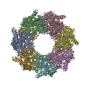
 マップデータ マップデータ | Volume contains GroEL complexed with two copies of non-native capsid protein gp23 | |||||||||
 試料 試料 |
| |||||||||
 キーワード キーワード | chaperonin / bacteriophage T4 / capsid protein gp23 / GroEL | |||||||||
| 機能・相同性 | Capsid protein, T4-like bacteriophage-like / protein binding / Chaperonin Cpn60/GroEL 機能・相同性情報 機能・相同性情報 | |||||||||
| 生物種 |   Enterobacteria phage T4 (ファージ) Enterobacteria phage T4 (ファージ) | |||||||||
| 手法 | 単粒子再構成法 / クライオ電子顕微鏡法 / ネガティブ染色法 / 解像度: 10.3 Å | |||||||||
 データ登録者 データ登録者 | Clare DK / Bakkes PJ / van Heerikhuizen H / van der Vies SM / Saibil HR | |||||||||
 引用 引用 |  ジャーナル: Nature / 年: 2009 ジャーナル: Nature / 年: 2009タイトル: Chaperonin complex with a newly folded protein encapsulated in the folding chamber. 著者: D K Clare / P J Bakkes / H van Heerikhuizen / S M van der Vies / H R Saibil /  要旨: A subset of essential cellular proteins requires the assistance of chaperonins (in Escherichia coli, GroEL and GroES), double-ring complexes in which the two rings act alternately to bind, ...A subset of essential cellular proteins requires the assistance of chaperonins (in Escherichia coli, GroEL and GroES), double-ring complexes in which the two rings act alternately to bind, encapsulate and fold a wide range of nascent or stress-denatured proteins. This process starts by the trapping of a substrate protein on hydrophobic surfaces in the central cavity of a GroEL ring. Then, binding of ATP and co-chaperonin GroES to that ring ejects the non-native protein from its binding sites, through forced unfolding or other major conformational changes, and encloses it in a hydrophilic chamber for folding. ATP hydrolysis and subsequent ATP binding to the opposite ring trigger dissociation of the chamber and release of the substrate protein. The bacteriophage T4 requires its own version of GroES, gp31, which forms a taller folding chamber, to fold the major viral capsid protein gp23 (refs 16-20). Polypeptides are known to fold inside the chaperonin complex, but the conformation of an encapsulated protein has not previously been visualized. Here we present structures of gp23-chaperonin complexes, showing both the initial captured state and the final, close-to-native state with gp23 encapsulated in the folding chamber. Although the chamber is expanded, it is still barely large enough to contain the elongated gp23 monomer, explaining why the GroEL-GroES complex is not able to fold gp23 and showing how the chaperonin structure distorts to enclose a large, physiological substrate protein. | |||||||||
| 履歴 |
|
- 構造の表示
構造の表示
| ムービー |
 ムービービューア ムービービューア |
|---|---|
| 構造ビューア | EMマップ:  SurfView SurfView Molmil Molmil Jmol/JSmol Jmol/JSmol |
| 添付画像 |
- ダウンロードとリンク
-EMDBアーカイブ
| マップデータ |  emd_1545.map.gz emd_1545.map.gz | 211.2 KB |  EMDBマップデータ形式 EMDBマップデータ形式 | |
|---|---|---|---|---|
| ヘッダ (付随情報) |  emd-1545-v30.xml emd-1545-v30.xml emd-1545.xml emd-1545.xml | 11.8 KB 11.8 KB | 表示 表示 |  EMDBヘッダ EMDBヘッダ |
| 画像 |  1545.gif 1545.gif | 51.7 KB | ||
| アーカイブディレクトリ |  http://ftp.pdbj.org/pub/emdb/structures/EMD-1545 http://ftp.pdbj.org/pub/emdb/structures/EMD-1545 ftp://ftp.pdbj.org/pub/emdb/structures/EMD-1545 ftp://ftp.pdbj.org/pub/emdb/structures/EMD-1545 | HTTPS FTP |
-検証レポート
| 文書・要旨 |  emd_1545_validation.pdf.gz emd_1545_validation.pdf.gz | 212.7 KB | 表示 |  EMDB検証レポート EMDB検証レポート |
|---|---|---|---|---|
| 文書・詳細版 |  emd_1545_full_validation.pdf.gz emd_1545_full_validation.pdf.gz | 211.8 KB | 表示 | |
| XML形式データ |  emd_1545_validation.xml.gz emd_1545_validation.xml.gz | 5.2 KB | 表示 | |
| アーカイブディレクトリ |  https://ftp.pdbj.org/pub/emdb/validation_reports/EMD-1545 https://ftp.pdbj.org/pub/emdb/validation_reports/EMD-1545 ftp://ftp.pdbj.org/pub/emdb/validation_reports/EMD-1545 ftp://ftp.pdbj.org/pub/emdb/validation_reports/EMD-1545 | HTTPS FTP |
-関連構造データ
- リンク
リンク
| EMDBのページ |  EMDB (EBI/PDBe) / EMDB (EBI/PDBe) /  EMDataResource EMDataResource |
|---|
- マップ
マップ
| ファイル |  ダウンロード / ファイル: emd_1545.map.gz / 形式: CCP4 / 大きさ: 15.3 MB / タイプ: IMAGE STORED AS FLOATING POINT NUMBER (4 BYTES) ダウンロード / ファイル: emd_1545.map.gz / 形式: CCP4 / 大きさ: 15.3 MB / タイプ: IMAGE STORED AS FLOATING POINT NUMBER (4 BYTES) | ||||||||||||||||||||||||||||||||||||||||||||||||||||||||||||||||||||
|---|---|---|---|---|---|---|---|---|---|---|---|---|---|---|---|---|---|---|---|---|---|---|---|---|---|---|---|---|---|---|---|---|---|---|---|---|---|---|---|---|---|---|---|---|---|---|---|---|---|---|---|---|---|---|---|---|---|---|---|---|---|---|---|---|---|---|---|---|---|
| 注釈 | Volume contains GroEL complexed with two copies of non-native capsid protein gp23 | ||||||||||||||||||||||||||||||||||||||||||||||||||||||||||||||||||||
| 投影像・断面図 | 画像のコントロール
画像は Spider により作成 | ||||||||||||||||||||||||||||||||||||||||||||||||||||||||||||||||||||
| ボクセルのサイズ | X=Y=Z: 2.8 Å | ||||||||||||||||||||||||||||||||||||||||||||||||||||||||||||||||||||
| 密度 |
| ||||||||||||||||||||||||||||||||||||||||||||||||||||||||||||||||||||
| 対称性 | 空間群: 1 | ||||||||||||||||||||||||||||||||||||||||||||||||||||||||||||||||||||
| 詳細 | EMDB XML:
CCP4マップ ヘッダ情報:
| ||||||||||||||||||||||||||||||||||||||||||||||||||||||||||||||||||||
-添付データ
- 試料の構成要素
-全体 : GroEL-gp23(2)
| 全体 | 名称: GroEL-gp23(2) |
|---|---|
| 要素 |
|
-超分子 #1000: GroEL-gp23(2)
| 超分子 | 名称: GroEL-gp23(2) / タイプ: sample / ID: 1000 詳細: gp23 was denatured in UREA and mixed with GroEL containing buffer 集合状態: tetradecamer of GroEL bound to one copy of denatured gp23 Number unique components: 2 |
|---|---|
| 分子量 | 実験値: 900 KDa / 理論値: 900 KDa / 手法: sequence |
-分子 #1: GroEL
| 分子 | 名称: GroEL / タイプ: protein_or_peptide / ID: 1 / Name.synonym: GroEL / コピー数: 1 / 集合状態: tetradecamer / 組換発現: Yes |
|---|---|
| 由来(天然) | 生物種:  |
| 分子量 | 実験値: 56 KDa / 理論値: 56 KDa |
| 組換発現 | 生物種:  |
| 配列 | GO: protein binding / InterPro: Chaperonin Cpn60/GroEL |
-分子 #2: gp23
| 分子 | 名称: gp23 / タイプ: protein_or_peptide / ID: 2 / Name.synonym: Major capsid protein / コピー数: 2 / 集合状態: monomer / 組換発現: Yes |
|---|---|
| 由来(天然) | 生物種:  Enterobacteria phage T4 (ファージ) / 別称: Bacteriophage T4 / 細胞中の位置: cytoplasmic Enterobacteria phage T4 (ファージ) / 別称: Bacteriophage T4 / 細胞中の位置: cytoplasmic |
| 分子量 | 実験値: 56 KDa / 理論値: 56 KDa |
| 組換発現 | 生物種:  |
| 配列 | InterPro: Capsid protein, T4-like bacteriophage-like |
-実験情報
-構造解析
| 手法 | ネガティブ染色法, クライオ電子顕微鏡法 |
|---|---|
 解析 解析 | 単粒子再構成法 |
| 試料の集合状態 | particle |
- 試料調製
試料調製
| 濃度 | 1 mg/mL |
|---|---|
| 緩衝液 | pH: 7.5 / 詳細: 40 mM Tris-HCl pH 7.5, 10 mM KCl, 10 mM MgCl2 |
| 染色 | タイプ: NEGATIVE / 詳細: Cryo |
| グリッド | 詳細: 400 mesh R2/2 c-flat grids |
| 凍結 | 凍結剤: ETHANE / チャンバー内温度: 100 K / 装置: HOMEMADE PLUNGER / 詳細: Vitrification instrument: manual plunger / 手法: blot for 2-3 seconds before plunging |
- 電子顕微鏡法
| 顕微鏡 | FEI TECNAI F20 |
|---|---|
| 温度 | 平均: 100 K |
| アライメント法 | Legacy - 非点収差: corrected at 150,00 times |
| 日付 | 2006年6月1日 |
| 撮影 | カテゴリ: FILM / フィルム・検出器のモデル: KODAK SO-163 FILM / デジタル化 - スキャナー: ZEISS SCAI / デジタル化 - サンプリング間隔: 1.4 µm / 実像数: 150 / 平均電子線量: 15 e/Å2 / Od range: 0.5 / ビット/ピクセル: 8 |
| 電子線 | 加速電圧: 200 kV / 電子線源:  FIELD EMISSION GUN FIELD EMISSION GUN |
| 電子光学系 | 倍率(補正後): 50000 / 照射モード: FLOOD BEAM / 撮影モード: BRIGHT FIELD / Cs: 2 mm / 最大 デフォーカス(公称値): 3.0 µm / 最小 デフォーカス(公称値): 1.0 µm / 倍率(公称値): 50000 |
| 試料ステージ | 試料ホルダー: side entry / 試料ホルダーモデル: GATAN LIQUID NITROGEN |
| 実験機器 |  モデル: Tecnai F20 / 画像提供: FEI Company |
- 画像解析
画像解析
| 詳細 | No symmetry was applied |
|---|---|
| CTF補正 | 詳細: Each particle |
| 最終 再構成 | 想定した対称性 - 点群: C1 (非対称) / アルゴリズム: OTHER / 解像度のタイプ: BY AUTHOR / 解像度: 10.3 Å / 解像度の算出法: FSC 0.5 CUT-OFF / ソフトウェア - 名称: SPIDER / 詳細: No symmetry was applied to the reconstruction / 使用した粒子像数: 6200 |
| 最終 角度割当 | 詳細: SPIDER:theta 78-90 degrees, phi 0-360 degrees. Mirror option was on. |
-原子モデル構築 1
| 初期モデル | PDB ID: |
|---|---|
| ソフトウェア | 名称: URO |
| 詳細 | Protocol: Individual domains fitted as rigid bodies. The domains were separately fitted in URO |
| 精密化 | 空間: RECIPROCAL / プロトコル: RIGID BODY FIT 当てはまり具合の基準: cross-correlation coefficient |
 ムービー
ムービー コントローラー


 Z (Sec.)
 X (Row.)
 Y (Col.)


