+ データを開く
- 基本情報
基本情報
| 登録情報 | データベース: EMDB / ID: EMD-1541 | |||||||||
|---|---|---|---|---|---|---|---|---|---|---|
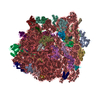
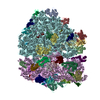
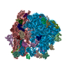
| タイトル | Visualization of the hybrid state of tRNA binding promoted by spontaneous ratcheting of the ribosome | |||||||||
 マップデータ マップデータ | Hybrid configuration pre-translocational ribosome | |||||||||
 試料 試料 |
| |||||||||
 キーワード キーワード | Hybrid state tRNA configuration / pre-translocational ribosome / spontaneous ratchet motion | |||||||||
| 生物種 |  | |||||||||
| 手法 | 単粒子再構成法 / クライオ電子顕微鏡法 / ネガティブ染色法 / 解像度: 8.9 Å | |||||||||
 データ登録者 データ登録者 | Agirrezabala X / Lei J / Brunelle JL / Ortiz-Meoz RF / Green R / Frank J | |||||||||
 引用 引用 |  ジャーナル: Mol Cell / 年: 2008 ジャーナル: Mol Cell / 年: 2008タイトル: Visualization of the hybrid state of tRNA binding promoted by spontaneous ratcheting of the ribosome. 著者: Xabier Agirrezabala / Jianlin Lei / Julie L Brunelle / Rodrigo F Ortiz-Meoz / Rachel Green / Joachim Frank /  要旨: A crucial step in translation is the translocation of tRNAs through the ribosome. In the transition from one canonical site to the other, the tRNAs acquire intermediate configurations, so-called ...A crucial step in translation is the translocation of tRNAs through the ribosome. In the transition from one canonical site to the other, the tRNAs acquire intermediate configurations, so-called hybrid states. At this stage, the small subunit is rotated with respect to the large subunit, and the anticodon stem loops reside in the A and P sites of the small subunit, while the acceptor ends interact with the P and E sites of the large subunit. In this work, by means of cryo-EM and particle classification procedures, we visualize the hybrid state of both A/P and P/E tRNAs in an authentic factor-free ribosome complex during translocation. In addition, we show how the repositioning of the tRNAs goes hand in hand with the change in the interplay between S13, L1 stalk, L5, H68, H69, and H38 that is caused by the ratcheting of the small subunit. | |||||||||
| 履歴 |
|
- 構造の表示
構造の表示
| ムービー |
 ムービービューア ムービービューア |
|---|---|
| 構造ビューア | EMマップ:  SurfView SurfView Molmil Molmil Jmol/JSmol Jmol/JSmol |
| 添付画像 |
- ダウンロードとリンク
-EMDBアーカイブ
| マップデータ |  emd_1541.map.gz emd_1541.map.gz | 54.6 MB |  EMDBマップデータ形式 EMDBマップデータ形式 | |
|---|---|---|---|---|
| ヘッダ (付随情報) |  emd-1541-v30.xml emd-1541-v30.xml emd-1541.xml emd-1541.xml | 11.1 KB 11.1 KB | 表示 表示 |  EMDBヘッダ EMDBヘッダ |
| 画像 |  1541.gif 1541.gif | 1.3 MB | ||
| アーカイブディレクトリ |  http://ftp.pdbj.org/pub/emdb/structures/EMD-1541 http://ftp.pdbj.org/pub/emdb/structures/EMD-1541 ftp://ftp.pdbj.org/pub/emdb/structures/EMD-1541 ftp://ftp.pdbj.org/pub/emdb/structures/EMD-1541 | HTTPS FTP |
-検証レポート
| 文書・要旨 |  emd_1541_validation.pdf.gz emd_1541_validation.pdf.gz | 242 KB | 表示 |  EMDB検証レポート EMDB検証レポート |
|---|---|---|---|---|
| 文書・詳細版 |  emd_1541_full_validation.pdf.gz emd_1541_full_validation.pdf.gz | 241.2 KB | 表示 | |
| XML形式データ |  emd_1541_validation.xml.gz emd_1541_validation.xml.gz | 6.2 KB | 表示 | |
| アーカイブディレクトリ |  https://ftp.pdbj.org/pub/emdb/validation_reports/EMD-1541 https://ftp.pdbj.org/pub/emdb/validation_reports/EMD-1541 ftp://ftp.pdbj.org/pub/emdb/validation_reports/EMD-1541 ftp://ftp.pdbj.org/pub/emdb/validation_reports/EMD-1541 | HTTPS FTP |
-関連構造データ
- リンク
リンク
| EMDBのページ |  EMDB (EBI/PDBe) / EMDB (EBI/PDBe) /  EMDataResource EMDataResource |
|---|---|
| 「今月の分子」の関連する項目 |
- マップ
マップ
| ファイル |  ダウンロード / ファイル: emd_1541.map.gz / 形式: CCP4 / 大きさ: 58.2 MB / タイプ: IMAGE STORED AS FLOATING POINT NUMBER (4 BYTES) ダウンロード / ファイル: emd_1541.map.gz / 形式: CCP4 / 大きさ: 58.2 MB / タイプ: IMAGE STORED AS FLOATING POINT NUMBER (4 BYTES) | ||||||||||||||||||||||||||||||||||||||||||||||||||||||||||||||||||||
|---|---|---|---|---|---|---|---|---|---|---|---|---|---|---|---|---|---|---|---|---|---|---|---|---|---|---|---|---|---|---|---|---|---|---|---|---|---|---|---|---|---|---|---|---|---|---|---|---|---|---|---|---|---|---|---|---|---|---|---|---|---|---|---|---|---|---|---|---|---|
| 注釈 | Hybrid configuration pre-translocational ribosome | ||||||||||||||||||||||||||||||||||||||||||||||||||||||||||||||||||||
| 投影像・断面図 | 画像のコントロール
画像は Spider により作成 | ||||||||||||||||||||||||||||||||||||||||||||||||||||||||||||||||||||
| ボクセルのサイズ | X=Y=Z: 1.5 Å | ||||||||||||||||||||||||||||||||||||||||||||||||||||||||||||||||||||
| 密度 |
| ||||||||||||||||||||||||||||||||||||||||||||||||||||||||||||||||||||
| 対称性 | 空間群: 1 | ||||||||||||||||||||||||||||||||||||||||||||||||||||||||||||||||||||
| 詳細 | EMDB XML:
CCP4マップ ヘッダ情報:
| ||||||||||||||||||||||||||||||||||||||||||||||||||||||||||||||||||||
-添付データ
- 試料の構成要素
-全体 : Hybrid state pre-translocational ribosome
| 全体 | 名称: Hybrid state pre-translocational ribosome |
|---|---|
| 要素 |
|
-超分子 #1000: Hybrid state pre-translocational ribosome
| 超分子 | 名称: Hybrid state pre-translocational ribosome / タイプ: sample / ID: 1000 / 詳細: HiFi buffer, 3.5 mM Mg2 / 集合状態: Monomeric / Number unique components: 3 |
|---|---|
| 分子量 | 実験値: 2.6 MDa / 理論値: 2.6 MDa / 手法: Sedimentation |
-超分子 #1: 70S prokaryote ribosome
| 超分子 | 名称: 70S prokaryote ribosome / タイプ: complex / ID: 1 / Name.synonym: Ribosome / 詳細: Pre-translocational ribosome / 組換発現: No / Ribosome-details: ribosome-prokaryote: ALL |
|---|---|
| 由来(天然) | 生物種:  |
| 分子量 | 実験値: 2.6 MDa / 理論値: 2.6 MDa |
-実験情報
-構造解析
| 手法 | ネガティブ染色法, クライオ電子顕微鏡法 |
|---|---|
 解析 解析 | 単粒子再構成法 |
| 試料の集合状態 | particle |
- 試料調製
試料調製
| 濃度 | 0.07 mg/mL |
|---|---|
| 緩衝液 | pH: 7.5 詳細: HiFi (50 mM Tris-HCl pH 7.5, 70mM NH4Cl, 30 mM KCl, 3.5 mM MgCl2, 0.5 mM spermidine, 8mM putrescine, 2 mM DTT) |
| 染色 | タイプ: NEGATIVE / 詳細: Cryo-sample |
| グリッド | 詳細: Quantifoil 2/4 |
| 凍結 | 凍結剤: NITROGEN / チャンバー内湿度: 90 % / チャンバー内温度: 80 K / 装置: OTHER / 詳細: Vitrification instrument: Vitrobot / 手法: Blot for 6 seconds before plunging |
- 電子顕微鏡法
| 顕微鏡 | FEI TECNAI F30 |
|---|---|
| 温度 | 最低: 80.7 K / 最高: 80.7 K / 平均: 80.7 K |
| アライメント法 | Legacy - 非点収差: Objective corrected at 100,000 times magnification |
| 特殊光学系 | エネルギーフィルター - 名称: No energy filter |
| 詳細 | Automated data collection system AutoEMation (CCD mag. 100000x) |
| 日付 | 2007年4月15日 |
| 撮影 | カテゴリ: CCD フィルム・検出器のモデル: TVIPS TEMCAM-F415 (4k x 4k) 平均電子線量: 25 e/Å2 詳細: Automated data collection system AutoEMation (CCD mag. 100000x) TVIPS TemCam-F415 |
| Tilt angle min | 0 |
| Tilt angle max | 0 |
| 電子線 | 加速電圧: 300 kV / 電子線源:  FIELD EMISSION GUN FIELD EMISSION GUN |
| 電子光学系 | 倍率(補正後): 58269 / 照射モード: FLOOD BEAM / 撮影モード: BRIGHT FIELD / Cs: 2.26 mm / 最大 デフォーカス(公称値): 4.0 µm / 最小 デフォーカス(公称値): 1.2 µm / 倍率(公称値): 59000 |
| 試料ステージ | 試料ホルダー: Cartridge / 試料ホルダーモデル: OTHER |
| 実験機器 |  モデル: Tecnai F30 / 画像提供: FEI Company |
 ムービー
ムービー コントローラー


 Z (Sec.)
 Y (Row.)
 X (Col.)


