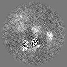
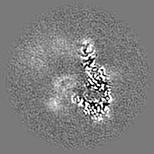
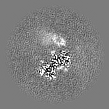
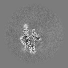
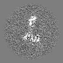
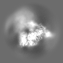
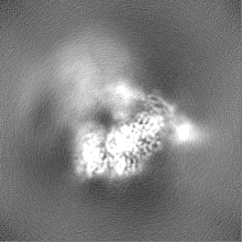
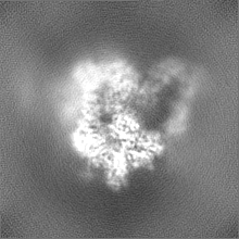
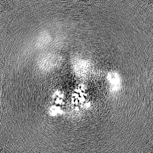
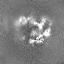
Journal: Nat Commun / Year: 2022 Title: Structure of the proteolytic enzyme PAPP-A with the endogenous inhibitor stanniocalcin-2 reveals its inhibitory mechanism. Authors: Sara Dam Kobberø / Michael Gajhede / Osman Asghar Mirza / Søren Kløverpris / Troels Rønn Kjær / Jakob Hauge Mikkelsen / Thomas Boesen / Claus Oxvig / Abstract: The metzincin metalloproteinase PAPP-A plays a key role in the regulation of insulin-like growth factor (IGF) signaling by specific cleavage of inhibitory IGF binding proteins (IGFBPs). Using single- ...The metzincin metalloproteinase PAPP-A plays a key role in the regulation of insulin-like growth factor (IGF) signaling by specific cleavage of inhibitory IGF binding proteins (IGFBPs). Using single-particle cryo-electron microscopy (cryo-EM), we here report the structure of PAPP-A in complex with its endogenous inhibitor, stanniocalcin-2 (STC2), neither of which have been reported before. The highest resolution (3.1 Å) was obtained for the STC2 subunit and the N-terminal approximately 1000 residues of the PAPP-A subunit. The 500 kDa 2:2 PAPP-A·STC2 complex is a flexible multidomain ensemble with numerous interdomain contacts. In particular, a specific disulfide bond between the subunits of STC2 and PAPP-A prevents dissociation, and interactions between STC2 and a module located in the very C-terminal end of the PAPP-A subunit prevent binding of its main substrate, IGFBP-4. While devoid of activity towards IGFBP-4, the active site cleft of the catalytic domain is accessible in the inhibited PAPP-A·STC2 complex, as shown by its ability to hydrolyze a synthetic peptide derived from IGFBP-4. Relevant to multiple human pathologies, this unusual mechanism of proteolytic inhibition may support the development of specific pharmaceutical agents, by which IGF signaling can be indirectly modulated.
Entire : Partial PAPP-A dimer in complex with subunit of its endogenous in...
Entire
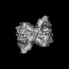
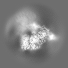
Name: Partial PAPP-A dimer in complex with subunit of its endogenous inhibitor STC2 dimer
Components
Complex: Partial PAPP-A dimer in complex with subunit of its endogenous inhibitor STC2 dimer
Complex: PAPP-A
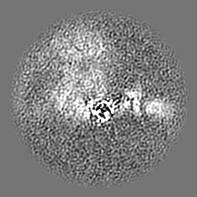
Complex: N-terminal part of PAPP-A
Protein or peptide: Pappalysin-1
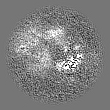
Complex: C-terminal part of PAPP-A
Complex: Subunit of Stanniocalcin-2
Protein or peptide: Stanniocalcin-2
Ligand: 2-acetamido-2-deoxy-beta-D-glucopyranose
Ligand: ZINC ION
Ligand: CALCIUM ION
+
Supramolecule #1: Partial PAPP-A dimer in complex with subunit of its endogenous in...
Supramolecule
Name: Partial PAPP-A dimer in complex with subunit of its endogenous inhibitor STC2 dimer type: complex / ID: 1 / Parent: 0 / Macromolecule list: #1-#2 Details: Inhibited proteolytic complex generated by harvest of serum media, purifying on a nickel column followed by negative affinity purification and size-exclusion chromatography.
Source (natural)
Organism: Homo sapiens (human) / Tissue: ubiquitous / Location in cell: extracellular
Molecular weight
Theoretical: 500 KDa
+
Supramolecule #2: PAPP-A
Supramolecule
Name: PAPP-A / type: complex / ID: 2 / Parent: 1 / Macromolecule list: #1 / Details: Parts of both PAPP-A subunits
Source (natural)
Organism: Homo sapiens (human) / Tissue: ubiquitous / Location in cell: extracellular
+
Supramolecule #3: Subunit of Stanniocalcin-2
Supramolecule
Name: Subunit of Stanniocalcin-2 / type: complex / ID: 3 / Parent: 1 / Macromolecule list: #2 / Details: One subunit of the STC2 dimer
Source (natural)
Organism: Homo sapiens (human) / Tissue: ubiquitous / Location in cell: extracellular
+
Supramolecule #4: N-terminal part of PAPP-A
Supramolecule
Name: N-terminal part of PAPP-A / type: complex / ID: 4 / Parent: 2 / Macromolecule list: #1 Details: The first approximately 1000 residues of a PAPP-A subunit
Source (natural)
Organism: Homo sapiens (human) / Tissue: ubiquitous / Location in cell: extracellular
+
Supramolecule #5: C-terminal part of PAPP-A
Supramolecule
Name: C-terminal part of PAPP-A / type: complex / ID: 5 / Parent: 2 / Macromolecule list: #1 / Details: The C-terminal 240 residues of one PAPP-A subunit
Source (natural)
Organism: Homo sapiens (human) / Tissue: ubiquitous / Location in cell: extracellular
+
Macromolecule #1: Pappalysin-1
Macromolecule
Name: Pappalysin-1 / type: protein_or_peptide / ID: 1 / Number of copies: 2 / Enantiomer: LEVO / EC number: pappalysin-1
Source (natural)
Organism: Homo sapiens (human) / Cell: extracellular
Name: ZINC ION / type: ligand / ID: 4 / Number of copies: 1 / Formula: ZN
Molecular weight
Theoretical: 65.409 Da
+
Macromolecule #5: CALCIUM ION
Macromolecule
Name: CALCIUM ION / type: ligand / ID: 5 / Number of copies: 8 / Formula: CA
Molecular weight
Theoretical: 40.078 Da
-
Experimental details
-
Structure determination
Method
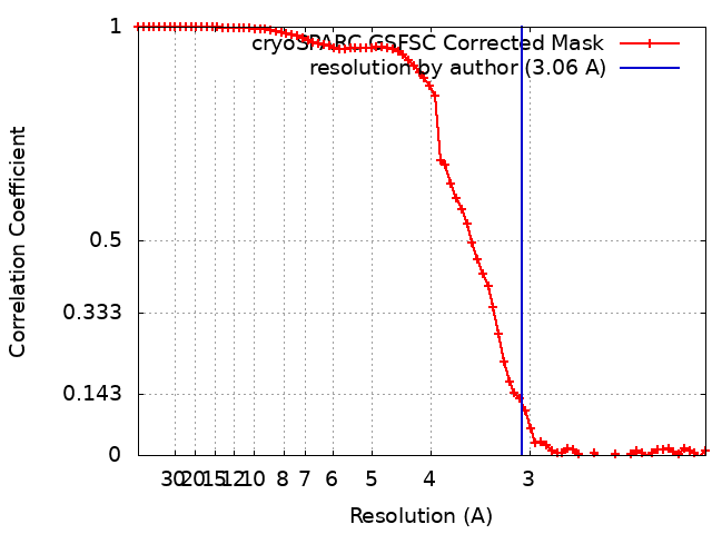
cryo EM
Processing
single particle reconstruction
Aggregation state
particle
-
Sample preparation
Concentration
0.6 mg/mL
Buffer
pH: 7.4 Component:
Concentration
Formula
Name
20.0 mM
Hepes
Hepes
100.0 mM
NaCl
NaCL
1.0 mM
CaCl
CaCl
Details: Hepes buffer, 20 mM Hepes pH 7.4 100 mM NaCl, 1 mM CaCl
Grid
Model: C-flat-2/2 / Material: COPPER / Mesh: 400 / Support film - Material: CARBON / Support film - topology: HOLEY / Pretreatment - Type: GLOW DISCHARGE / Pretreatment - Time: 45 sec.
Vitrification
Cryogen name: ETHANE / Chamber humidity: 90 % / Chamber temperature: 277.15 K / Instrument: LEICA EM GP / Details: 4 s.
-
Electron microscopy
Microscope
FEI TITAN KRIOS
Image recording
#0 - Image recording ID: 1 / #0 - Film or detector model: GATAN K3 BIOQUANTUM (6k x 4k) / #0 - Number grids imaged: 1 / #0 - Number real images: 32144 / #0 - Average exposure time: 0.91 sec. / #0 - Average electron dose: 59.0 e/Å2 / #1 - Image recording ID: 2 / #1 - Film or detector model: GATAN K3 BIOQUANTUM (6k x 4k) / #1 - Number grids imaged: 1 / #1 - Number real images: 10060 / #1 - Average exposure time: 0.8 sec. / #1 - Average electron dose: 58.0 e/Å2
Electron beam
Acceleration voltage: 300 kV / Electron source: FIELD EMISSION GUN
In the structure databanks used in Yorodumi, some data are registered as the other names, "COVID-19 virus" and "2019-nCoV". Here are the details of the virus and the list of structure data.
Jan 31, 2019. EMDB accession codes are about to change! (news from PDBe EMDB page)
EMDB accession codes are about to change! (news from PDBe EMDB page)
The allocation of 4 digits for EMDB accession codes will soon come to an end. Whilst these codes will remain in use, new EMDB accession codes will include an additional digit and will expand incrementally as the available range of codes is exhausted. The current 4-digit format prefixed with “EMD-” (i.e. EMD-XXXX) will advance to a 5-digit format (i.e. EMD-XXXXX), and so on. It is currently estimated that the 4-digit codes will be depleted around Spring 2019, at which point the 5-digit format will come into force.
The EM Navigator/Yorodumi systems omit the EMD- prefix.
Related info.:Q: What is EMD? / ID/Accession-code notation in Yorodumi/EM Navigator
Yorodumi is a browser for structure data from EMDB, PDB, SASBDB, etc.
This page is also the successor to EM Navigator detail page, and also detail information page/front-end page for Omokage search.
The word "yorodu" (or yorozu) is an old Japanese word meaning "ten thousand". "mi" (miru) is to see.
Related info.:EMDB / PDB / SASBDB / Comparison of 3 databanks / Yorodumi Search / Aug 31, 2016. New EM Navigator & Yorodumi / Yorodumi Papers / Jmol/JSmol / Function and homology information / Changes in new EM Navigator and Yorodumi
 Movie
Movie Controller


 Open data
 Basic information
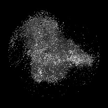
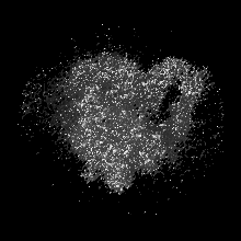
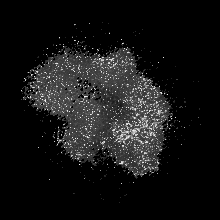
 Map data
 Sample
 Keywords
 Function and homology information
 Homo sapiens (human)
 Authors
 Denmark, 1 items
  Citation
 Structure visualization
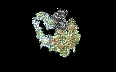
 Downloads & links
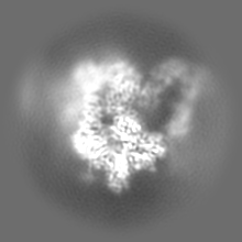
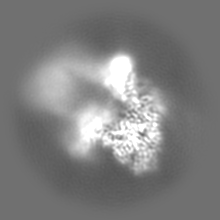
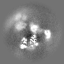
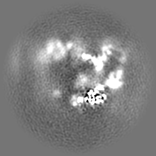
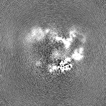
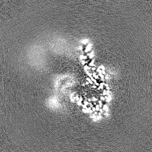
 emd_15220.png
 http://ftp.pdbj.org/pub/emdb/structures/EMD-15220


 Z (Sec.)
 Y (Row.)
 X (Col.)


 Sample components
 Processing
 Electron microscopy
 FIELD EMISSION GUN