 Movie
Movie Controller

[English] 日本語
 Yorodumi
- EMDB-1459: Partitivirus structure reveals a 120-subunit, helix-rich capsid w... -
+ Open data

- Basic information
| Entry | Database: EMDB / ID: EMD-1459 | |||||||||
|---|---|---|---|---|---|---|---|---|---|---|
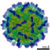
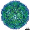
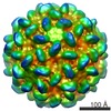
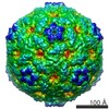
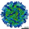
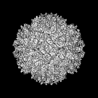
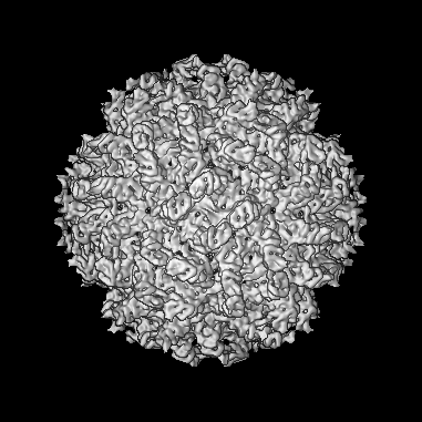
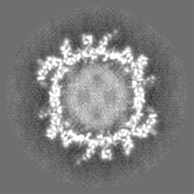
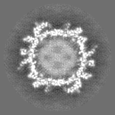
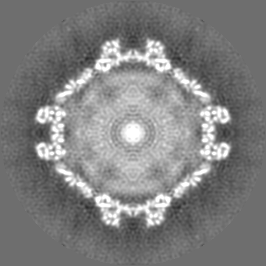
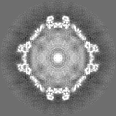
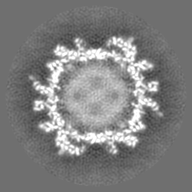
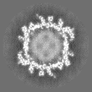
| Title | Partitivirus structure reveals a 120-subunit, helix-rich capsid with distinctive surface arches formed by quasisymmetric coat-protein dimers. | |||||||||
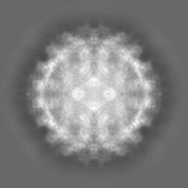
 Map data Map data | This is the final map calculated | |||||||||
 Sample Sample |
| |||||||||
| Biological species |  Penicillium stoloniferum virus S Penicillium stoloniferum virus S | |||||||||
| Method | single particle reconstruction / cryo EM / Resolution: 7.3 Å | |||||||||
 Authors Authors | Ochoa WF / Havens WM / Sinkovits RS / Nibert ML / Ghabrial SA / Baker TS | |||||||||
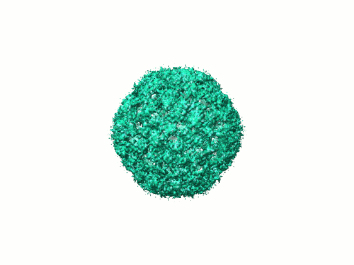
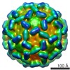
 Citation Citation | Journal: Structure / Year: 2008 Title: Partitivirus structure reveals a 120-subunit, helix-rich capsid with distinctive surface arches formed by quasisymmetric coat-protein dimers. Authors: Wendy F Ochoa / Wendy M Havens / Robert S Sinkovits / Max L Nibert / Said A Ghabrial / Timothy S Baker /  Abstract: Two distinct partitiviruses, Penicillium stoloniferum viruses S and F, can be isolated from the fungus Penicillium stoloniferum. The bisegmented dsRNA genomes of these viruses are separately packaged ...Two distinct partitiviruses, Penicillium stoloniferum viruses S and F, can be isolated from the fungus Penicillium stoloniferum. The bisegmented dsRNA genomes of these viruses are separately packaged in icosahedral capsids containing 120 coat-protein subunits. We used transmission electron cryomicroscopy and three-dimensional image reconstruction to determine the structure of Penicillium stoloniferum virus S at 7.3 A resolution. The capsid, approximately 350 A in outer diameter, contains 12 pentons, each of which is topped by five arched protrusions. Each of these protrusions is, in turn, formed by a quasisymmetric dimer of coat protein, for a total of 60 such dimers per particle. The density map shows numerous tubular features, characteristic of alpha helices and consistent with secondary structure predictions for the coat protein. This three-dimensional structure of a virus from the family Partitiviridae exhibits both similarities to and differences from the so-called "T = 2" capsids of other dsRNA viruses. | |||||||||
| History |
|
- Structure visualization
| Movie |
 Movie viewer Movie viewer |
|---|---|
| Structure viewer | EM map: SurfViewMolmilJmol/JSmol |
| Supplemental images |
- Downloads & links
-EMDB archive
| Map data | emd_1459.map.gz | 38.7 MB | EMDB map data format | |
|---|---|---|---|---|
| Header (meta data) | emd-1459-v30.xmlemd-1459.xml | 6.5 KB 6.5 KB | Display Display | EMDB header |
| Images |  1459.gif 1459.gif | 36.7 KB | ||
| Archive directory |  http://ftp.pdbj.org/pub/emdb/structures/EMD-1459ftp://ftp.pdbj.org/pub/emdb/structures/EMD-1459 http://ftp.pdbj.org/pub/emdb/structures/EMD-1459ftp://ftp.pdbj.org/pub/emdb/structures/EMD-1459 | HTTPS FTP |
-Related structure data
| Similar structure data |
|---|
-Links
| EMDB pages | EMDB (EBI/PDBe) / EMDataResource |
|---|
-Map
| File | Download / File: emd_1459.map.gz / Format: CCP4 / Size: 206 MB / Type: IMAGE STORED AS FLOATING POINT NUMBER (4 BYTES) | ||||||||||||||||||||||||||||||||||||||||||||||||||||||||||||||||||||
|---|---|---|---|---|---|---|---|---|---|---|---|---|---|---|---|---|---|---|---|---|---|---|---|---|---|---|---|---|---|---|---|---|---|---|---|---|---|---|---|---|---|---|---|---|---|---|---|---|---|---|---|---|---|---|---|---|---|---|---|---|---|---|---|---|---|---|---|---|---|
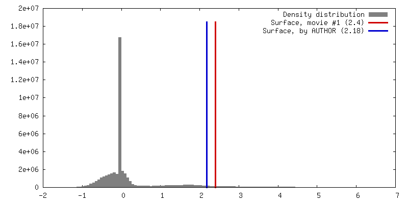
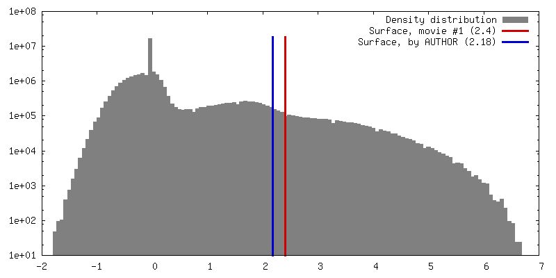
| Annotation | This is the final map calculated | ||||||||||||||||||||||||||||||||||||||||||||||||||||||||||||||||||||
| Projections & slices | Image control
Images are generated by Spider. | ||||||||||||||||||||||||||||||||||||||||||||||||||||||||||||||||||||
| Voxel size | X=Y=Z: 1.23 Å | ||||||||||||||||||||||||||||||||||||||||||||||||||||||||||||||||||||
| Density |
| ||||||||||||||||||||||||||||||||||||||||||||||||||||||||||||||||||||
| Symmetry | Space group: 1 | ||||||||||||||||||||||||||||||||||||||||||||||||||||||||||||||||||||
| Details | EMDB XML:
CCP4 map header:
| ||||||||||||||||||||||||||||||||||||||||||||||||||||||||||||||||||||
 Z (Sec.)
 Y (Row.)
 X (Col.)


-Supplemental data
- Sample components
-Entire : PsV-S
| Entire | Name: PsV-S |
|---|---|
| Components |
|
-Supramolecule #1000: PsV-S
| Supramolecule | Name: PsV-S / type: sample / ID: 1000 / Number unique components: 1 |
|---|
-Supramolecule #1: Penicillium stoloniferum virus S
| Supramolecule | Name: Penicillium stoloniferum virus S / type: virus / ID: 1 / Name.synonym: PsV-S / NCBI-ID: 216371 / Sci species name: Penicillium stoloniferum virus S / Virus type: VIRION / Virus isolate: SPECIES / Virus enveloped: No / Virus empty: No / Syn species name: PsV-S |
|---|---|
| Host (natural) | Organism:  Penicillium Stoloniferum (fungus) / synonym: FUNGI Penicillium Stoloniferum (fungus) / synonym: FUNGI |
| Virus shell | Shell ID: 1 / Name: PsV-S / Diameter: 35 Å / T number (triangulation number): 2 |
-Experimental details
-Structure determination
| Method | cryo EM |
|---|---|
 Processing Processing | single particle reconstruction |
| Aggregation state | particle |
-Sample preparation
| Vitrification | Cryogen name: ETHANE |
|---|
- Electron microscopy
| Microscope | FEI TECNAI SPHERA |
|---|---|
| Electron beam | Acceleration voltage: 200 kV / Electron source:  FIELD EMISSION GUN FIELD EMISSION GUN |
| Electron optics | Illumination mode: SPOT SCAN / Imaging mode: OTHER |
| Sample stage | Specimen holder: Polara / Specimen holder model: GATAN LIQUID NITROGEN |
-Image processing
| Final reconstruction | Applied symmetry - Point group: I (icosahedral) / Resolution.type: BY AUTHOR / Resolution: 7.3 Å / Resolution method: FSC 0.5 CUT-OFF |
|---|
