ムービー
ムービー コントローラー


+ データを開く

- 基本情報
基本情報
| 登録情報 | データベース: EMDB / ID: EMD-1439 | |||||||||
|---|---|---|---|---|---|---|---|---|---|---|
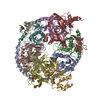
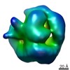
| タイトル | Architecture of the yeast Rrp44 exosome complex suggests routes of RNA recruitment for 3' end processing. | |||||||||
 マップデータ マップデータ | This is the 3D map of yeast exosome core complex | |||||||||
 試料 試料 |
| |||||||||
| 生物種 |  | |||||||||
| 手法 | 単粒子再構成法 / クライオ電子顕微鏡法 / ネガティブ染色法 / 解像度: 23.0 Å | |||||||||
 データ登録者 データ登録者 | Wang H-W / Wang J / Ding F / Callahan K / Bratkowski MA / Butler JS / Nogales E / Ke A | |||||||||
 引用 引用 | ジャーナル: Proc Natl Acad Sci U S A / 年: 2007 タイトル: Architecture of the yeast Rrp44 exosome complex suggests routes of RNA recruitment for 3' end processing. 著者: Hong-Wei Wang / Jianjun Wang / Fang Ding / Kevin Callahan / Matthew A Bratkowski / J Scott Butler / Eva Nogales / Ailong Ke /  要旨: The eukaryotic core exosome (CE) is a conserved nine-subunit protein complex important for 3' end trimming and degradation of RNA. In yeast, the Rrp44 protein constitutively associates with the CE ...The eukaryotic core exosome (CE) is a conserved nine-subunit protein complex important for 3' end trimming and degradation of RNA. In yeast, the Rrp44 protein constitutively associates with the CE and provides the sole source of processive 3'-to-5' exoribonuclease activity. Here we present EM reconstructions of the core and Rrp44-bound exosome complexes. The two-lobed Rrp44 protein binds to the RNase PH domain side of the exosome and buttresses the bottom of the exosome-processing chamber. The Rrp44 C-terminal body part containing an RNase II-type active site is anchored to the exosome through a conserved set of interactions mainly to the Rrp45 and Rrp43 subunit, whereas the Rrp44 N-terminal head part is anchored to the Rrp41 subunit and may function as a roadblock to restrict access of RNA to the active site in the body region. The Rrp44-exosome (RE) architecture suggests an active site sequestration mechanism for strict control of 3' exoribonuclease activity in the RE complex. | |||||||||
| 履歴 |
|
- 構造の表示
構造の表示
| ムービー |
 ムービービューア ムービービューア |
|---|---|
| 構造ビューア | EMマップ: SurfViewMolmilJmol/JSmol |
| 添付画像 |
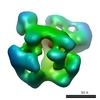
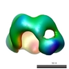
 UCSF Chimera
- ダウンロードとリンク
-EMDBアーカイブ
| マップデータ | emd_1439.map.gz | 1.2 MB | EMDBマップデータ形式 | |
|---|---|---|---|---|
| ヘッダ (付随情報) | emd-1439-v30.xmlemd-1439.xml | 15.9 KB 15.9 KB | 表示 表示 | EMDBヘッダ |
| 画像 | 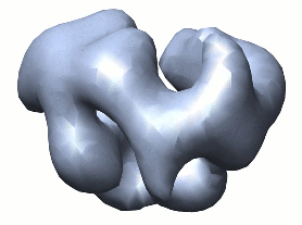 1439.gif 1439.gif | 30.6 KB | ||
| アーカイブディレクトリ |  http://ftp.pdbj.org/pub/emdb/structures/EMD-1439ftp://ftp.pdbj.org/pub/emdb/structures/EMD-1439 http://ftp.pdbj.org/pub/emdb/structures/EMD-1439ftp://ftp.pdbj.org/pub/emdb/structures/EMD-1439 | HTTPS FTP |
-検証レポート
| 文書・要旨 | emd_1439_validation.pdf.gz | 198.7 KB | 表示 | EMDB検証レポート |
|---|---|---|---|---|
| 文書・詳細版 | emd_1439_full_validation.pdf.gz | 197.9 KB | 表示 | |
| XML形式データ | emd_1439_validation.xml.gz | 5.2 KB | 表示 | |
| アーカイブディレクトリ | https://ftp.pdbj.org/pub/emdb/validation_reports/EMD-1439ftp://ftp.pdbj.org/pub/emdb/validation_reports/EMD-1439 | HTTPS FTP |
-関連構造データ


-リンク
| EMDBのページ | EMDB (EBI/PDBe) / EMDataResource |
|---|
-マップ
| ファイル | ダウンロード / ファイル: emd_1439.map.gz / 形式: CCP4 / 大きさ: 1.4 MB / タイプ: IMAGE STORED AS FLOATING POINT NUMBER (4 BYTES) | ||||||||||||||||||||||||||||||||||||||||||||||||||||||||||||||||||||
|---|---|---|---|---|---|---|---|---|---|---|---|---|---|---|---|---|---|---|---|---|---|---|---|---|---|---|---|---|---|---|---|---|---|---|---|---|---|---|---|---|---|---|---|---|---|---|---|---|---|---|---|---|---|---|---|---|---|---|---|---|---|---|---|---|---|---|---|---|---|
| 注釈 | This is the 3D map of yeast exosome core complex | ||||||||||||||||||||||||||||||||||||||||||||||||||||||||||||||||||||
| 投影像・断面図 | 画像のコントロール
画像は Spider により作成 | ||||||||||||||||||||||||||||||||||||||||||||||||||||||||||||||||||||
| ボクセルのサイズ | X=Y=Z: 5.18 Å | ||||||||||||||||||||||||||||||||||||||||||||||||||||||||||||||||||||
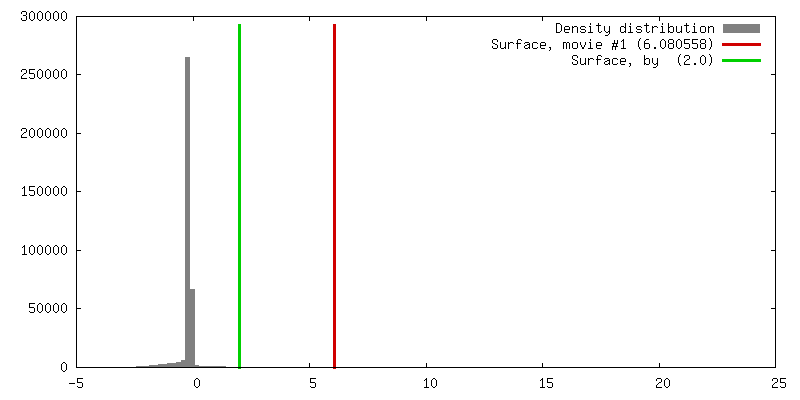
| 密度 |
| ||||||||||||||||||||||||||||||||||||||||||||||||||||||||||||||||||||
| 対称性 | 空間群: 1 | ||||||||||||||||||||||||||||||||||||||||||||||||||||||||||||||||||||
| 詳細 | EMDB XML:
CCP4マップ ヘッダ情報:
| ||||||||||||||||||||||||||||||||||||||||||||||||||||||||||||||||||||
 Z (Sec.)
 Y (Row.)
 X (Col.)


-添付データ
- 試料の構成要素
+全体 : exosome core complex
+超分子 #1000: exosome core complex
+分子 #1: Rrp43
+分子 #2: Rrp4
+分子 #3: Cls4
+分子 #4: Rrp45
+分子 #5: Rrp46-TAP
+分子 #6: Rrp41
+分子 #7: Rrp42
+分子 #8: Mtr3
+分子 #9: Rrp40
-実験情報
-構造解析
| 手法 | ネガティブ染色法, クライオ電子顕微鏡法 |
|---|---|
 解析 解析 | 単粒子再構成法 |
| 試料の集合状態 | particle |
-試料調製
| 濃度 | 0.03 mg/mL |
|---|---|
| 緩衝液 | pH: 7.5 詳細: 25 mM Tris-HCl, 50 mM NaCl, 2 mM DTT, and 10 uM ZnCl2 |
| 染色 | タイプ: NEGATIVE 詳細: Four microliters of the protein solution was negatively stained with 2% uranyl formate solution between two thin layers of carbon on a copper grid by using the sandwich method. |
| グリッド | 詳細: 400 mesh copper grid |
| 凍結 | 凍結剤: ETHANE |
- 電子顕微鏡法
| 顕微鏡 | FEI TECNAI 12 |
|---|---|
| 温度 | 平均: 300 K |
| アライメント法 | Legacy - 非点収差: objective lens astigmatism was corrected at 100,000 times magnification |
| 詳細 | Low dose mode was used for taking pictures |
| 日付 | 2006年8月1日 |
| 撮影 | カテゴリ: FILM / フィルム・検出器のモデル: KODAK SO-163 FILM / デジタル化 - スキャナー: OTHER / デジタル化 - サンプリング間隔: 12.7 µm / 実像数: 50 / 平均電子線量: 20 e/Å2 詳細: The micrographs were scanned on a Nikon Super Coolscan 8000 scanner Od range: 1.4 / ビット/ピクセル: 14 |
| Tilt angle min | 0 |
| Tilt angle max | 0 |
| 電子線 | 加速電圧: 120 kV / 電子線源: LAB6 |
| 電子光学系 | 倍率(補正後): 49000 / 照射モード: FLOOD BEAM / 撮影モード: BRIGHT FIELD / Cs: 6.6 mm / 最大 デフォーカス(公称値): 0.9 µm / 最小 デフォーカス(公称値): 0.7 µm / 倍率(公称値): 49000 |
| 試料ステージ | 試料ホルダー: normal single-tilt holder / 試料ホルダーモデル: OTHER |
-画像解析
| 最終 再構成 | 想定した対称性 - 点群: C1 (非対称) / アルゴリズム: OTHER / 解像度のタイプ: BY AUTHOR / 解像度: 23.0 Å / 解像度の算出法: FSC 0.5 CUT-OFF / ソフトウェア - 名称: SPIDER / 詳細: Final map were calculated from all the particles. / 使用した粒子像数: 2980 |
|---|---|
| 最終 角度割当 | 詳細: SPIDER: theta 90 degrees, phi 90 degrees |
| 最終 2次元分類 | クラス数: 50 |
-原子モデル構築 1
| 初期モデル | PDB ID: |
|---|---|
| ソフトウェア | 名称: Situs |
| 詳細 | Protocol: Rigid Body. The docking was performed by automatic rigid body docking in Situs. |
| 精密化 | 空間: REAL / プロトコル: RIGID BODY FIT |