 Movie
Movie Controller

[English] 日本語
 Yorodumi
- EMDB-1225: Loading a ring: structure of the Bacillus subtilis DnaB protein, ... -
+ Open data

- Basic information
| Entry | Database: EMDB / ID: EMD-1225 | |||||||||
|---|---|---|---|---|---|---|---|---|---|---|
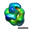
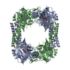
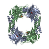
| Title | Loading a ring: structure of the Bacillus subtilis DnaB protein, a co-loader of the replicative helicase. | |||||||||
 Map data Map data | Bacillus subtilis DnaB | |||||||||
 Sample Sample |
| |||||||||
| Function / homology | :  Function and homology information Function and homology information | |||||||||
| Biological species |  | |||||||||
| Method | single particle reconstruction / negative staining / Resolution: 24.0 Å | |||||||||
 Authors Authors | Nunez-Ramirez R / Velten M / Rivas G / Polard P / Carazo JM / Donate LE | |||||||||
 Citation Citation | Journal: J Mol Biol / Year: 2007 Title: Loading a ring: structure of the Bacillus subtilis DnaB protein, a co-loader of the replicative helicase. Authors: Rafael Núñez-Ramírez / Marion Velten / Germán Rivas / Patrice Polard / José María Carazo / Luis Enrique Donate /  Abstract: Loading of the ring-shaped replicative helicase is a critical step in the initiation of DNA replication. Bacillus subtilis has adopted a two-protein strategy to load its hexameric replicative ...Loading of the ring-shaped replicative helicase is a critical step in the initiation of DNA replication. Bacillus subtilis has adopted a two-protein strategy to load its hexameric replicative helicase: DnaB and DnaI interact with the helicase and mediate its delivery onto DNA. We present here the 3D electron microscopy structure of the DnaB protein, along with a detailed analysis of both its oligomeric state and its domain organization. DnaB is organized as an asymmetric tetramer that is comprised of two stacked components, one arranged as a closed collar and the other as an open sigma shape. Intriguingly, the 3D map of DnaB exhibits an overall architecture similar to the structure of the Escherichia coli gamma-complex, the loader of the ring-shaped processivity factor. We propose a model whereby each DnaB monomer participates in both stacked components of the tetramer and displays a different overall shape. This asymmetric quaternary organization could be a general feature of ring loaders. | |||||||||
| History |
|
- Structure visualization
| Movie |
Movie viewer |
|---|---|
| Structure viewer | EM map: SurfViewMolmilJmol/JSmol |
| Supplemental images |
 UCSF Chimera
- Downloads & links
-EMDB archive
| Map data | emd_1225.map.gz | 942.6 KB | EMDB map data format | |
|---|---|---|---|---|
| Header (meta data) | emd-1225-v30.xmlemd-1225.xml | 8.7 KB 8.7 KB | Display Display | EMDB header |
| Images |  1225.gif 1225.gif | 6.7 KB | ||
| Archive directory |  http://ftp.pdbj.org/pub/emdb/structures/EMD-1225ftp://ftp.pdbj.org/pub/emdb/structures/EMD-1225 http://ftp.pdbj.org/pub/emdb/structures/EMD-1225ftp://ftp.pdbj.org/pub/emdb/structures/EMD-1225 | HTTPS FTP |
-Related structure data
| Similar structure data |
|---|


-Links
| EMDB pages | EMDB (EBI/PDBe) / EMDataResource |
|---|
-Map
| File | Download / File: emd_1225.map.gz / Format: CCP4 / Size: 1001 KB / Type: IMAGE STORED AS FLOATING POINT NUMBER (4 BYTES) | ||||||||||||||||||||||||||||||||||||||||||||||||||||||||||||||||||||
|---|---|---|---|---|---|---|---|---|---|---|---|---|---|---|---|---|---|---|---|---|---|---|---|---|---|---|---|---|---|---|---|---|---|---|---|---|---|---|---|---|---|---|---|---|---|---|---|---|---|---|---|---|---|---|---|---|---|---|---|---|---|---|---|---|---|---|---|---|---|
| Annotation | Bacillus subtilis DnaB | ||||||||||||||||||||||||||||||||||||||||||||||||||||||||||||||||||||
| Projections & slices | Image control
Images are generated by Spider. | ||||||||||||||||||||||||||||||||||||||||||||||||||||||||||||||||||||
| Voxel size | X=Y=Z: 4 Å | ||||||||||||||||||||||||||||||||||||||||||||||||||||||||||||||||||||
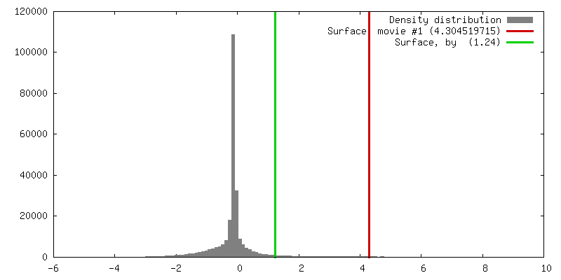
| Density |
| ||||||||||||||||||||||||||||||||||||||||||||||||||||||||||||||||||||
| Symmetry | Space group: 1 | ||||||||||||||||||||||||||||||||||||||||||||||||||||||||||||||||||||
| Details | EMDB XML:
CCP4 map header:
| ||||||||||||||||||||||||||||||||||||||||||||||||||||||||||||||||||||
 Z (Sec.)
 Y (Row.)
 X (Col.)


-Supplemental data
- Sample components
-Entire : Bacillus subtilis DnaB
| Entire | Name: Bacillus subtilis DnaB |
|---|---|
| Components |
|
-Supramolecule #1000: Bacillus subtilis DnaB
| Supramolecule | Name: Bacillus subtilis DnaB / type: sample / ID: 1000 / Oligomeric state: Homotetramer / Number unique components: 1 |
|---|---|
| Molecular weight | Experimental: 200 KDa / Theoretical: 200 KDa Method: Sedimentation equilibrium by analytical ultracentrifugation |
-Macromolecule #1: DnaB
| Macromolecule | Name: DnaB / type: protein_or_peptide / ID: 1 / Number of copies: 4 / Oligomeric state: Tetramer / Recombinant expression: Yes |
|---|---|
| Source (natural) | Organism: |
| Molecular weight | Experimental: 200 KDa / Theoretical: 200 KDa |
| Recombinant expression | Organism: |
| Sequence | InterPro: INTERPRO: IPR010833 |
-Experimental details
-Structure determination
| Method | negative staining |
|---|---|
 Processing Processing | single particle reconstruction |
| Aggregation state | particle |
-Sample preparation
| Concentration | 0.088 mg/mL |
|---|---|
| Buffer | pH: 7.5 / Details: 50mM Tris-HCl, 50mM NaCl, 2mM MgCl2 and 1mM DTT |
| Staining | Type: NEGATIVE Details: Grids with adsorbed protein stained on 2% uranyl acetate for 1 minute |
| Grid | Details: collodion/carbon coated 400 mesh copper grid |
| Vitrification | Cryogen name: NONE |
- Electron microscopy
| Microscope | JEOL 2000EX |
|---|---|
| Alignment procedure | Legacy - Astigmatism: objective lens astigmatism was corrected at 100,000 times magnification |
| Image recording | Category: FILM / Film or detector model: KODAK SO-163 FILM / Digitization - Scanner: ZEISS SCAI / Digitization - Sampling interval: 21 µm / Number real images: 20 / Average electron dose: 20 e/Å2 / Bits/pixel: 8 |
| Electron beam | Acceleration voltage: 80 kV / Electron source: TUNGSTEN HAIRPIN |
| Electron optics | Illumination mode: OTHER / Imaging mode: BRIGHT FIELD / Nominal magnification: 60000 |
| Sample stage | Specimen holder: Eucentric / Specimen holder model: OTHER |
-Image processing
| Final reconstruction | Applied symmetry - Point group: C1 (asymmetric) / Algorithm: OTHER / Resolution.type: BY AUTHOR / Resolution: 24.0 Å / Resolution method: FSC 0.5 CUT-OFF / Software - Name: Xmipp and EMAN / Number images used: 8352 |
|---|
