 Movie
Movie Controller

[English] 日本語
 Yorodumi
- EMDB-13694: Substrate-engaged mycobacterial Proteasome-associated ATPase - fo... -
+ Open data

- Basic information
| Entry | Database: EMDB / ID: EMD-13694 | |||||||||
|---|---|---|---|---|---|---|---|---|---|---|
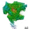
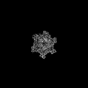
| Title | Substrate-engaged mycobacterial Proteasome-associated ATPase - focused 3D refinement (state A) | |||||||||
 Map data Map data | Substrate-engaged mycobacterial proteasome activator bound to 20S CP in state A (focused 3D refinement) | |||||||||
 Sample Sample |
| |||||||||
 Keywords Keywords | AAA motor / ATPAse / mycobacterium / proteasome activator / 20S CP / CYTOSOLIC PROTEIN | |||||||||
| Function / homology |  Function and homology information Function and homology informationprotein pupylation / proteasome-activating nucleotidase complex / proteasome binding / proteasomal protein catabolic process / modification-dependent protein catabolic process / protein tag activity / ATP hydrolysis activity / ATP binding Similarity search - Function | |||||||||
| Biological species |   Mycobacterium tuberculosis (bacteria) Mycobacterium tuberculosis (bacteria) | |||||||||
| Method | single particle reconstruction / cryo EM / Resolution: 3.8 Å | |||||||||
 Authors Authors | Jomaa A / Kavalchuk M | |||||||||
| Funding support |  Switzerland, 1 items Switzerland, 1 items
| |||||||||
 Citation Citation | Journal: Nat Commun / Year: 2022 Title: Structural basis of prokaryotic ubiquitin-like protein engagement and translocation by the mycobacterial Mpa-proteasome complex. Authors: Mikhail Kavalchuk / Ahmad Jomaa / Andreas U Müller / Eilika Weber-Ban / Abstract: Proteasomes are present in eukaryotes, archaea and Actinobacteria, including the human pathogen Mycobacterium tuberculosis, where proteasomal degradation supports persistence inside the host. In ...Proteasomes are present in eukaryotes, archaea and Actinobacteria, including the human pathogen Mycobacterium tuberculosis, where proteasomal degradation supports persistence inside the host. In mycobacteria and other members of Actinobacteria, prokaryotic ubiquitin-like protein (Pup) serves as a degradation tag post-translationally conjugated to target proteins for their recruitment to the mycobacterial proteasome ATPase (Mpa). Here, we use single-particle cryo-electron microscopy to determine the structure of Mpa in complex with the 20S core particle at an early stage of pupylated substrate recruitment, shedding light on the mechanism of substrate translocation. Two conformational states of Mpa show how substrate is translocated stepwise towards the degradation chamber of the proteasome core particle. We also demonstrate, in vitro and in vivo, the importance of a structural feature in Mpa that allows formation of alternating charge-complementary interactions with the proteasome resulting in radial, rail-guided movements during the ATPase conformational cycle. | |||||||||
| History |
|
- Structure visualization
| Movie |
Movie viewer |
|---|---|
| Structure viewer | EM map: SurfViewMolmilJmol/JSmol |
| Supplemental images |

- Downloads & links
-EMDB archive
| Map data | emd_13694.map.gz | 152.8 MB | EMDB map data format | |
|---|---|---|---|---|
| Header (meta data) | emd-13694-v30.xmlemd-13694.xml | 14.5 KB 14.5 KB | Display Display | EMDB header |
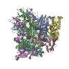
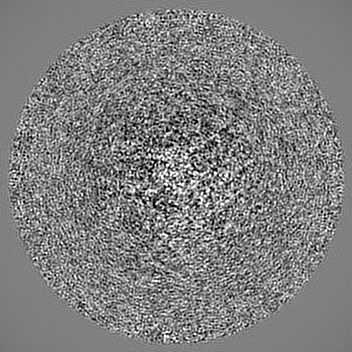
| Images | 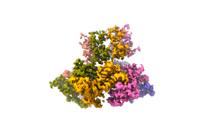 emd_13694.png emd_13694.png | 51 KB | ||
| Filedesc metadata | emd-13694.cif.gz | 6.5 KB | ||
| Archive directory |  http://ftp.pdbj.org/pub/emdb/structures/EMD-13694ftp://ftp.pdbj.org/pub/emdb/structures/EMD-13694 http://ftp.pdbj.org/pub/emdb/structures/EMD-13694ftp://ftp.pdbj.org/pub/emdb/structures/EMD-13694 | HTTPS FTP |
-Related structure data
| Related structure data |  7px9MC  7pxaC  7pxbC  7pxcC  7pxdC M: atomic model generated by this map C: citing same article ( |
|---|---|
| Similar structure data |


-Links
| EMDB pages | EMDB (EBI/PDBe) / EMDataResource |
|---|---|
| Related items in Molecule of the Month |


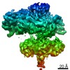
-Map
| File | Download / File: emd_13694.map.gz / Format: CCP4 / Size: 166.4 MB / Type: IMAGE STORED AS FLOATING POINT NUMBER (4 BYTES) | ||||||||||||||||||||||||||||||||||||||||||||||||||||||||||||||||||||
|---|---|---|---|---|---|---|---|---|---|---|---|---|---|---|---|---|---|---|---|---|---|---|---|---|---|---|---|---|---|---|---|---|---|---|---|---|---|---|---|---|---|---|---|---|---|---|---|---|---|---|---|---|---|---|---|---|---|---|---|---|---|---|---|---|---|---|---|---|---|
| Annotation | Substrate-engaged mycobacterial proteasome activator bound to 20S CP in state A (focused 3D refinement) | ||||||||||||||||||||||||||||||||||||||||||||||||||||||||||||||||||||
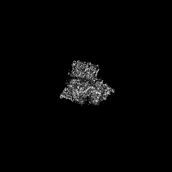
| Projections & slices | Image control
Images are generated by Spider. | ||||||||||||||||||||||||||||||||||||||||||||||||||||||||||||||||||||
| Voxel size | X=Y=Z: 1.222 Å | ||||||||||||||||||||||||||||||||||||||||||||||||||||||||||||||||||||
| Density |
| ||||||||||||||||||||||||||||||||||||||||||||||||||||||||||||||||||||
| Symmetry | Space group: 1 | ||||||||||||||||||||||||||||||||||||||||||||||||||||||||||||||||||||
| Details | EMDB XML:
CCP4 map header:
| ||||||||||||||||||||||||||||||||||||||||||||||||||||||||||||||||||||
 Z (Sec.)
 Y (Row.)
 X (Col.)


-Supplemental data
- Sample components
-Entire : Mycobacterial Proteasome-associated ATPase in complex with substr...
| Entire | Name: Mycobacterial Proteasome-associated ATPase in complex with substrate and open-gate 20SCP |
|---|---|
| Components |
|
-Supramolecule #1: Mycobacterial Proteasome-associated ATPase in complex with substr...
| Supramolecule | Name: Mycobacterial Proteasome-associated ATPase in complex with substrate and open-gate 20SCP type: complex / ID: 1 / Parent: 0 / Macromolecule list: #1-#2 |
|---|---|
| Source (natural) | Organism: Mycobacterium tuberculosis (bacteria) |
| Molecular weight | Theoretical: 1.1 MDa |
-Macromolecule #1: AAA ATPase forming ring-shaped complexes
| Macromolecule | Name: AAA ATPase forming ring-shaped complexes / type: protein_or_peptide / ID: 1 / Number of copies: 6 / Enantiomer: LEVO |
|---|---|
| Source (natural) | Organism: Mycobacterium tuberculosis (bacteria) |
| Molecular weight | Theoretical: 67.48793 KDa |
| Recombinant expression | Organism: |
| Sequence | String: MGESERSEAF GIPRDSPLSS GDAAELEQLR REAAVLREQL ENAVGSHAPT RSARDIHQLE ARIDSLAARN SKLMETLKEA RQQLLALRE EVDRLGQPPS GYGVLLATHD DDTVDVFTSG RKMRLTCSPN IDAASLKKGQ TVRLNEALTV VEAGTFEAVG E ISTLREIL ...String: MGESERSEAF GIPRDSPLSS GDAAELEQLR REAAVLREQL ENAVGSHAPT RSARDIHQLE ARIDSLAARN SKLMETLKEA RQQLLALRE EVDRLGQPPS GYGVLLATHD DDTVDVFTSG RKMRLTCSPN IDAASLKKGQ TVRLNEALTV VEAGTFEAVG E ISTLREIL ADGHRALVVG HADEERVVWL ADPLIAEDLP DGLPEALNDD TRPRKLRPGD SLLVDTKAGY AFERIPKAEV ED LVLEEVP DVSYADIGGL SRQIEQIRDA VELPFLHKEL YREYSLRPPK GVLLYGPPGC GKTLIAKAVA NSLAKKMAEV RGD DAHEAK SYFLNIKGPE LLNKFVGETE RHIRLIFQRA REKASEGTPV IVFFDEMDSI FRTRGTGVSS DVETTVVPQL LSEI DGVEG LENVIVIGAS NREDMIDPAI LRPGRLDVKI KIERPDAEAA QDIYSKYLTE FLPVHADDLA EFDGDRSACI KAMIE KVVD RMYAEIDDNR FLEVTYANGD KEVMYFKDFN SGAMIQNVVD RAKKNAIKSV LETGQPGLRI QHLLDSIVDE FAENED LPN TTNPDDWARI SGKKGERIVY IRTLVTGKSS SASRAIDTES NLGQYL UniProtKB: AAA ATPase forming ring-shaped complexes |
-Macromolecule #2: Prokaryotic ubiquitin-like protein Pup
| Macromolecule | Name: Prokaryotic ubiquitin-like protein Pup / type: protein_or_peptide / ID: 2 / Details: GS residues are left after tag cleavage / Number of copies: 1 / Enantiomer: LEVO |
|---|---|
| Source (natural) | Organism: Mycobacterium tuberculosis (bacteria) |
| Molecular weight | Theoretical: 7.095416 KDa |
| Recombinant expression | Organism: |
| Sequence | String: GSMAQEQTKR GGGGGDDDDI AGSTAAGQER REKLTEETDD LLDEIDDVLE ENAEDFVRAY VQKGGQ UniProtKB: Prokaryotic ubiquitin-like protein Pup |
-Macromolecule #3: ADENOSINE-5'-TRIPHOSPHATE
| Macromolecule | Name: ADENOSINE-5'-TRIPHOSPHATE / type: ligand / ID: 3 / Number of copies: 5 / Formula: ATP |
|---|---|
| Molecular weight | Theoretical: 507.181 Da |
| Chemical component information |  ChemComp-ATP: |
-Macromolecule #4: MAGNESIUM ION
| Macromolecule | Name: MAGNESIUM ION / type: ligand / ID: 4 / Number of copies: 5 / Formula: MG |
|---|---|
| Molecular weight | Theoretical: 24.305 Da |
-Macromolecule #5: ADENOSINE-5'-DIPHOSPHATE
| Macromolecule | Name: ADENOSINE-5'-DIPHOSPHATE / type: ligand / ID: 5 / Number of copies: 1 / Formula: ADP |
|---|---|
| Molecular weight | Theoretical: 427.201 Da |
| Chemical component information |  ChemComp-ADP: |
-Experimental details
-Structure determination
| Method | cryo EM |
|---|---|
 Processing Processing | single particle reconstruction |
| Aggregation state | particle |
-Sample preparation
| Buffer | pH: 7.5 |
|---|---|
| Grid | Model: Quantifoil R2/1 / Material: COPPER / Pretreatment - Type: GLOW DISCHARGE / Pretreatment - Time: 15 sec. |
| Vitrification | Cryogen name: ETHANE-PROPANE / Instrument: FEI VITROBOT MARK IV |
- Electron microscopy
| Microscope | FEI TITAN KRIOS |
|---|---|
| Image recording | Film or detector model: GATAN K3 (6k x 4k) / Average electron dose: 50.0 e/Å2 |
| Electron beam | Acceleration voltage: 300 kV / Electron source:  FIELD EMISSION GUN FIELD EMISSION GUN |
| Electron optics | Illumination mode: FLOOD BEAM / Imaging mode: BRIGHT FIELD / Nominal defocus max: 2.5 µm / Nominal defocus min: 1.5 µm / Nominal magnification: 105000 |
| Sample stage | Specimen holder model: FEI TITAN KRIOS AUTOGRID HOLDER / Cooling holder cryogen: NITROGEN |
| Experimental equipment |  Model: Titan Krios / Image courtesy: FEI Company |

